
Alveolar cleft reconstruction with vomerine bone: two surgical procedures in one step: a case series
 0
0 ,
, Abstract
Aim: Iliac crest (IC) is the most common source of bone for alveolar cleft repair, as it allows for harvesting a large amount of cancellous bone with a high rate of favorable outcomes. Its drawback is the donor-site morbidity. We propose a new technique for alveolar cleft grafting using vomerine bone (VB), which, through bone grafting, reconstructs the alveolus with the VB and simultaneously corrects the nasal septum deviation.
Method: We performed 18 alveolar reconstructions with VB in patients with a small bony defect and septal deviation, which would benefit from septoplasty. A matched control group with IC bone grafting was selected. Panoramic X-rays were used for vertical assessment of ossification with the Bergland scale and CT scans for the evaluation of the thickness of the grafted area in the VB and IC groups and compared with an independent samples T-test. A paired T-test compared angular measurements of the septal deviation pre- and post- vomerine grafting.
Results: All grafts healed uneventfully, with no complications at the donor site and respiratory function was improved. There was no statistically significant difference in ossification height between VB and IC, the alveolar thickness at the occlusal and middle third was higher with IC. Septal deviation was reduced significantly.
Conclusion: Alveolar graft from VB seems to be a viable alternative to IC in patients who present with a mild bony alveolar defect in addition to septal deviation, allowing combined procedures while reducing the morbidity of the donor site.
Keywords
INTRODUCTION
Alveolar cleft treatment is one of the most controversial topics in cleft lip and palate surgery. Many different techniques and timings have been proposed in order to obtain the best ossification of the alveolar cleft, ranging from primary periosteoplasty (PPP) to early secondary gingivoalveoloplasty (esGAP)[1], from primary bone grafting (PBG) to secondary bone grafting (SBG). The goal of the treatment is to supply bone for erupting teeth and periodontal support for the teeth adjacent to the cleft. SBG is a well-established technique to treat alveolar bone defects, according to the original description by Boyne and Sands[2]. The procedure is classified as early secondary if done between the ages of 2-5 years, intermediate secondary if done between the ages of 6-13 years in the late mixed dentition, or late secondary (also called tertiary) if done from adolescence onward, after the eruption of permanent teeth in the cleft area[3]. In our center, PBG was never performed, given the evidence of sagittal and especially vertical maxillary growth disturbances that occur after this procedure, as demonstrated in the Eurocleft study[4]. Alveolar bone grafting should be performed at the correct timing for optimal outcomes before the eruption of the permanent maxillary canine[5] or lateral incisor[6]. The timing considered ideal for grafting is when the unerupted lateral incisor root (canine if the lateral is missing) has reached one-half to two-thirds of development[7] or when the crowns of the erupting permanent teeth are covered by a thin shell of bone[8-9]. For late secondary cases, the procedure was done in patients who had a previous unsuccessful SBG or esGAP without a complete ossification, when the patient was ready to receive implant surgery or for orthodontic movement of the teeth in the cleft area. It is well known that late grafts do not achieve ideal alveolar height restoration as that occurs in earlier grafting, where bone remodeling is enhanced by the stimulating effect of the migrating teeth.
The goal of alveolar cleft grafting is to fill the defect with cancellous bone, which allows a faster healing process, and to reconstruct the nasal floor with cortical bone in order to restore the superior wall and resist the contraction of the overlying soft tissue as it heals. By resisting these contractile forces, the bone graft has a better chance to maintain its volume and potentially reduces the need for an additional graft. Many different donor sites have been proposed over time. IC is the most popular choice for SBG because it allows for harvesting a cortical segment to reconstruct the nasal floor and a large amount of cancellous bone to fill the defect[10].
Another source of membranous bone that has been utilized in alveolar cleft grafting is mandibular symphysis; harvesting from the mandible has the advantage of the use of the same operative field, no visible scar, and decreased postoperative pain[11]. The mandible can provide adequate bone volume for alveolar grafting even from the retromolar region[12].
Calvaria and tibia have been used by some authors as a source of cortical and cancellous bone[13]. Rib has also been utilized to close the alveolar cleft; however, it is considered to have limited indication due to its donor site morbidity, including visible scarring and pain. Rib grafts have also been criticized for the quality of regenerated bone with difficulties in orthodontic tooth movement[14]. In our Center, we traditionally perform bone graft harvesting cortical and cancellous bone from IC and it is still the gold standard.
We present a case series of alveolar reconstruction using vomerine bone. The idea behind the procedure was to couple the bone graft with the correction of the deviation of the nasal septum, solving at the same time respiratory problems that typically concern cleft patients and reconstructing the alveolar defect using the bone of the vomer. To our knowledge, this bone source has never been used before for alveolar reconstruction in growing patients.
METHODS
This study was conducted following the ethical principles outlined in the Declaration of Helsinki. Since 2015, at the Smile House in San Paolo Hospital of Milan, we have performed alveolar grafting with VB in all the UCLP patients with a clinically significant septal deviation when the alveolar bone defect is small enough to receive a complete reconstruction with this bone. A very small defect was not considered worthy of a major bone harvest, as IC.
The presence of a septal deviation was confirmed with a physical examination, with panoramic X-ray and CBCT. We deemed possible the reconstruction of defects that are no wider than 1 cm at the nasal floor and 2-3 mm at the lower alveolar level. Mucoperiosteal flaps are elevated on the palate and vestibular surfaces of the maxillary segments. Bone stumps of the alveolar cleft are exposed after reflection flaps. Nasal floor is released from the inferior attachments and elevated [Figures 1 and 2]. The excess tissue is trimmed in order to raise the floor as high as possible and then sutured in place. When construction of the watertight nasal floor is completed, the area is ready to receive the bone graft [Figures 3 and 4].
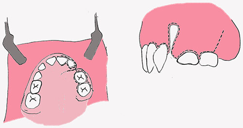
Figure 1. Schematic drawing of local condition at the time of bone graft.
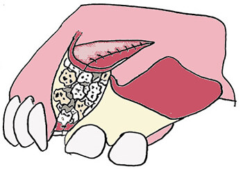
Figure 2. Schematic drawing of alveolar cleft after filling with bone.
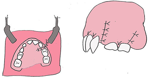
Figure 3. Schematic drawing with flaps sutured.
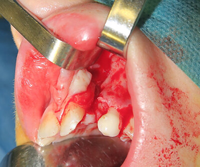
Figure 4. Intraoperative picture of alveolar cleft after elevation and closure of nasal floor.
Harvesting VB can be done with direct visualization or with endoscopic control, the same way septoplasty is performed. We used a hemitransfix incision in all cases according to the standard septoplasty technique. After a wide subpericondral-subperiostal dissection of the septum, in both cartilagineous and bony areas, we harvested the vomer keeping intact the perpendicular plate of the ethmoid bone [Figure 5]. The trick is to harvest bone fragments as wide as possible to reconstruct the alveolar walls with one-piece fragments. A single fragment of cortical bone is placed to reconstruct the nasal floor, and then small bone particles are densely packed into the cleft (same as done with cancellous bone when IC is the source of graft)
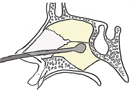
Figure 5. Schematic drawing of the area of septal dissection for access to vomerine bone and harvest through hemitransfix incision.
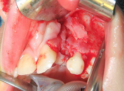
Figure 6. Intraoperative picture of alveolar cleft after filling inlay with vomerine bone.
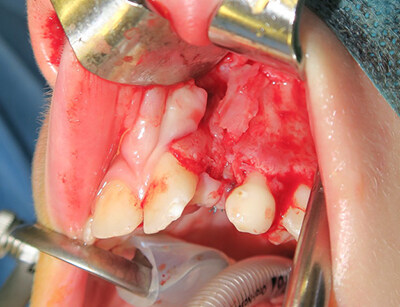
Figure 7. Intraoperative picture of alveolar cleft after coverage onlay with vomerine bone.
We performed 18 bone grafts with this technique, with a wide range in age of the patients (7.3-17.2 years, average 11.4). The sample included nonhomogenous cases, both unilateral complete cleft lip and palate patients and cleft lip and alveolus patients, previously treated in different centers. In detail, eight patients had received a primary treatment in our Center, with an all-in-one closure of lip, nose, soft and hard palate without any alveolar repair. Those patients had a primary cartilagineous septal repositioning.
Ten patients were treated in other centers. Six of them underwent a multi-stage closure of the cleft without any alveolar treatment, and four had already received surgery for alveolar restoration with a relatively poor outcome with only a small amount of bone bridging the cleft, therefore needing a bone augmentation of the existing bridge. No more information was available for patients coming from different centers than ours. We selected the patients who had a septal deviation with subsequent stenosis of the nasal fossa in order to improve nasal function. Of these patients, 16 had sufficient records to allow for the assessment of the quality of ossification.
A sample of patients subjected to traditional IC SBG was matched for age and cleft type and severity pre-grafting (no pre-graft clefts wider than 1 cm at the nasal floor and 2-3 mm at the lower alveolar level were considered) to be used as a control sample. The number of patients was too small to allow matching for gender or for cleft side.
Therefore, two samples were considered. The first sample consisted of 16 non-syndromic unilateral cleft lip and palate patients who underwent SBG with VB. They were treated according to the anatomical limits described by the senior surgeon (L.A.). The average age at the time of vomer SBG was of 11.4 ± 2.6 years. The second sample consisted of nine non-syndromic unilateral cleft lip and palate patients who underwent alveolar bone grafting from IC operated on by the same surgeon (L.A.). The sample was matched for preoperative width. The average age at the time of alveolar bone grafting was 10.4 ± 1.2 years.
We standardized the radiological follow-up of these patients with a panoramic X-Ray at 3 months, a panoramic X-Ray and CBCT at 1 year postoperatively. After a postop follow-up of approximately one year, we may presume there will be minimal change in the volume of the graft over the long term[15]. Evaluation on panoramic radiographs included two parameters: alveolar bridging and nasal ossification. The reliability of the use of panoramic radiographs instead of periapical films was demonstrated previously[16]. Alveolar bridging was assessed using a modified Bergland’s scoring system[5,16]. Type I (score 1): complete ossification of the cleft with normal alveolar reconstruction. Type II (score 2): interdental septal height more than three-quarters of the normal height. Type III (score 3): ossification less than three-quarters of the normal height. Type IV (score 4): ossification limited to a bridge or no ossification at all.
Nasal area ossification was given three different qualitative scores by the authors, as visible on the panoramic radiographs. Type I (score 1): flat symmetrical nasal floor. Type II (score 2): slightly asymmetrical notched nasal floor. Type III (score 3): severely asymmetrical notched nasal floor. Type IV (score 4): incomplete nasal floor[16].
Alveolar cleft thickness was also quantitatively evaluated on computed tomographic images and three-dimensional reconstructions of the maxilla. The quantitative analysis provided both absolute normalized measurements and ratios of the affected side versus the non-affected side. To compare cleft and non-cleft sides, the method from Meazzini et al. was adopted: a midline running through the center of the cervical vertebrae and the airways was drawn, and the thinnest area on the cleft side and its mirror section on the noncleft side was selected[17]. Panoramic radiograph for evaluation of septal deviation was already used with reliable results in a study for identification of the more suitable nostril for nasotracheal intubation[18].
Septal deviation pre- and post-grafting was assessed by calculating the angle between a line drawn through the septum and a line drawn across the infraorbital points (the lowest point of the infraorbital borders) [Figure 8]. Differences between ossification in IC and in VB grafting were compared with an independent sample T-test, setting P value at 0.05. Differences between septal deviation pre-surgery and post-VB grafting were compared with a Paired T-test, setting P value at 0.05.
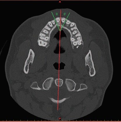
Figure 8. Method used to point out the areas of comparison between the cleft and non-cleft sides: red line, the midline passing through the cervical vertebra and the airway; green lines, the line passing through the thinnest section of the alveolar cleft and its specular line on the noncleft side (θ = θ′). Meazzini et al., Plast Reconstr Surg, 2016[17].
RESULTS
Two examiners repeated all measurements. The weighted kappa values, for inter-examiner reproducibility, demonstrated good agreement with an average weighted kappa coefficient of 0.88 (95%CI: 0.79-1.00) for Bergland ossification scores, almost perfect agreement with an average weighted kappa coefficient of
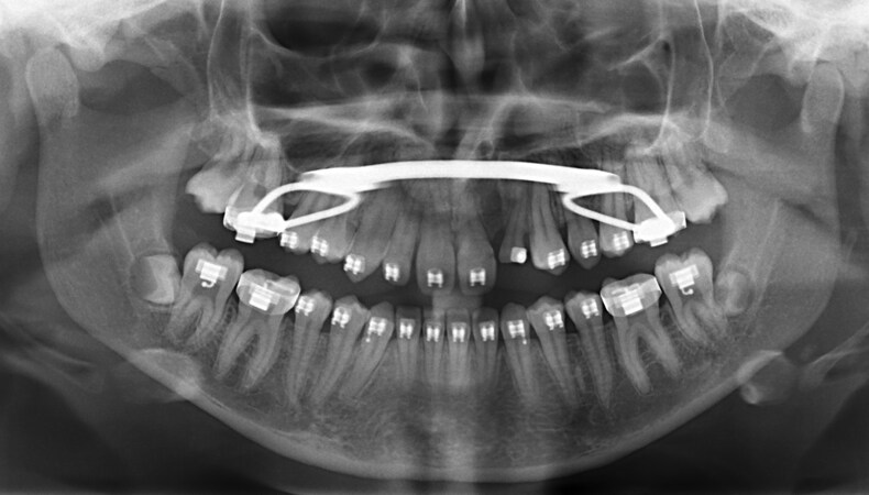
Figure 9. Patient M.S. Preoperative panoramic X-Ray, showing the alveolar area with a minimal bony defect in height and a moderate septal deviation.
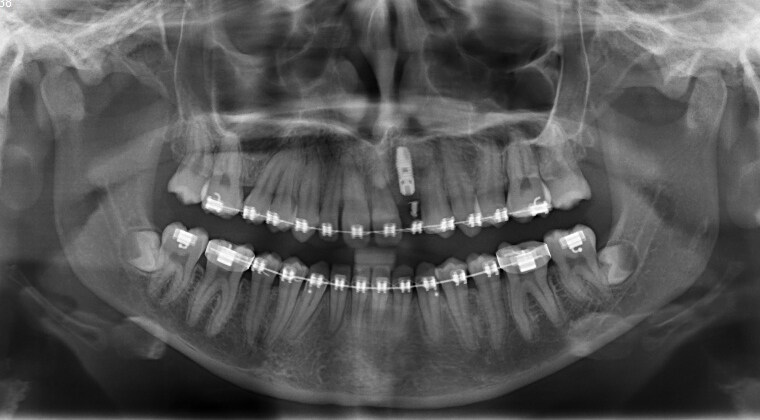
Figure 10. Patient M.S. Postoperative control, showing the restoration of a normal alveolar height, with subsequent implant rehabilitation, a symmetrical nasal floor and the correction of the septal deviation.
In a case with a narrow alveolar cleft and a severe septal deviation, bone graft with vomerine harvest allowed us to reach a good result with complete ossification of the alveolar cleft, normal height, slightly notched nasal floor, significant improvement of septal deviation viewed with orthopantomography
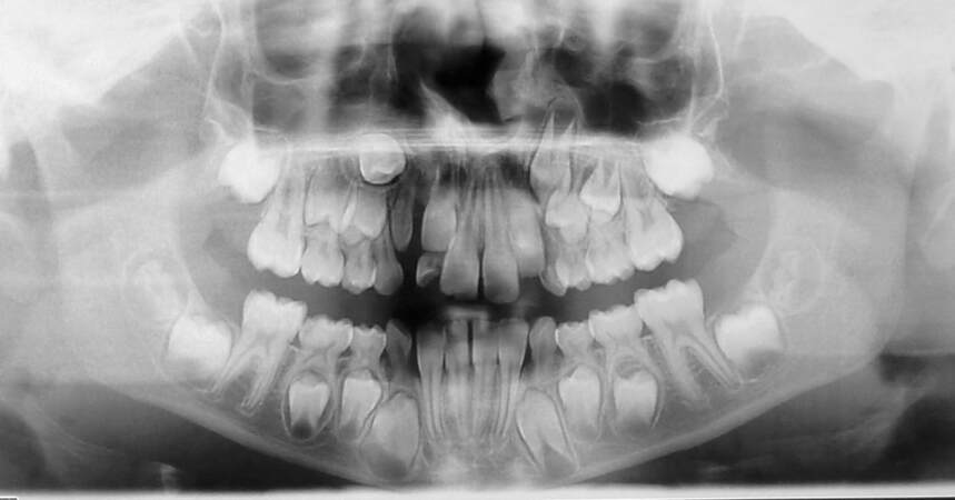
Figure 11. Patient D.G. Preoperative panoramic X-Ray: a narrow alveolar cleft is evident, as well as a severe septal deviation.
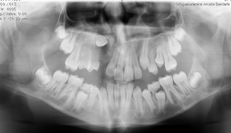
Figure 12. Patient D.G. Postoperative panoramic X-Ray, with complete ossification of the alveolar cleft, normal height, slightly notched nasal floor, significant improvement of septal deviation.
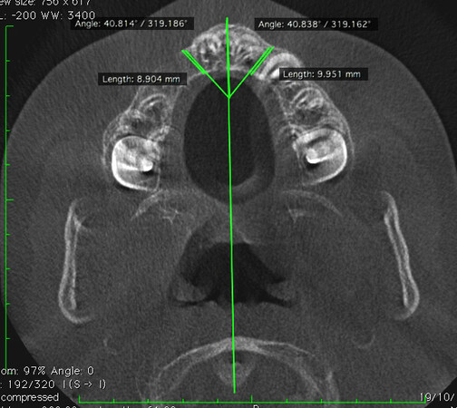
Figure 13. Patient D.G. (same as Figures 11 and 12): Axial scans showing degrees of alveolar thickness in the cleft area, compared to the non-cleft side, using the mirroring method described by Meazzini et al, 2016[17]. Ossification Type 1: bone thickness on the cleft side is 89% of the thickness on the noncleft side, classified as ideal.
Some cases healed with a less optimal result, rated as good ossification of the cleft (Type 2 of Meazzini’s classification), and bone thickness on the cleft side is 52 percent of the thickness on the non-cleft side [Figure 14].
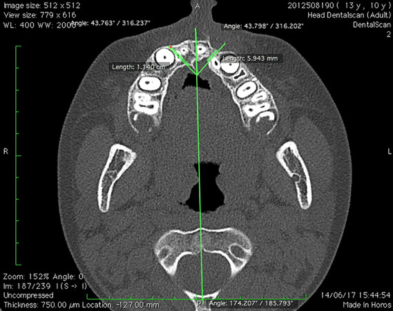
Figure 14. Patient G.A. Good ossification of the cleft (Type 2 of Meazzini’s classification), bone thickness on the cleft side is 52 percent of the thickness on the non-cleft side.
No differences were found between crest grafting and vomer grafting in Bergland’s modified classification of alveolar bone height or in Meazzini’s classification of nasal floor ossification [Table 1]. On the 3D CT evaluation, IC grafting allowed for a significantly thicker alveolar ridge at the most occlusal and middle third. There was no difference in the cranial third. The data regarding frequencies, means and standard deviations, together with the P values of the student T-test, are shown in Table 1.
Comparison between ossification after bone grafting with VB and IC. Alveolar height and thickness and nasal ossification are compared
| Bergland score panoramic X Ray | Nasal ossification | Alveolar ossification thickness on CT Ratio cleft/non cleft thickness | ||||
| 1: 100% 2: 75% 3: 50% 4: 0% | 1: flat 2: asymmetric 3: severe notch 4: no continuity | Occlusal third | Middle third | Cranial third | ||
| Vomer BG | X | 1.21 | 1.85 | 0.48 | 0.54 | 0.54 |
| sd | 0.43 | 0.47 | 0.29 | 0.21 | 0.20 | |
| Iliac crest BG | X | 1,67 | 1.77 | 0.75 | 0.67 | 0.34 |
| sd | 0.50 | 0.8 | 0.14 | 0.11 | 0.33 | |
| Independent samples T-test | 0.031 | 0.79 | 0.002** | 0.013* | 0.062 | |
All patients experienced an improvement in respiratory function, as reported in the clinical charts. The evaluation of septal deviation showed a substantial improvement of about 16 degrees relative to the infraorbital plane. The average preoperative deviation was 62.8 ± 17.3, while the postoperative deviation was 78.6 ± 9.8. The P value of the Paired T-test was 0.008. Vomerine BG was initially performed in patients that had a previous unsuccessful SBG or an esGAP without a complete ossification, in order to allow orthodontic tooth movement or eruption in the cleft area. We extended the indication to untouched alveolar clefts with a minimal bony defect. The width of the defects was no wider than 1 cm at the nasal floor and 2-3 mm at the lower alveolar level.
DISCUSSION
The goal of alveolar cleft grafting is to fill the defect with cancellous bone, which allows a faster healing process, and to reconstruct the nasal floor with cortical bone in order to restore the nasal floor and resist the contraction of the overlying soft tissue as it heals.
The IC is worldwide considered the first choice as a donor site, because it provides a large amount of cancellous and cortical bone for reconstructing the volume. Iliac bone harvest is a very easy and safe procedure, requires special attention in preserving the growth cartilage and avoiding cutaneous nerve injury, and has two major drawbacks, the pain during postoperative mobilization with delayed ambulation and the scar, which can be very visible.
Cranial bone is an alternative that allows the surgeon to avoid visible scars. While calvaria provides a large amount of cortical dense bone and has a very easy postoperative recovery, the operative time is much longer and may have major complications (intracranial penetration with hemorrhage, infections, and dural tear)[17-19]. Tibia has also been used as a source of cortico-cancellous bone harvested with a trephine. It is a painless procedure with minimal discomfort in walking for a few days and has a smaller scar compared with IC. Care, though, has to be taken to avoid damage to the tibial articulating margin and to a correct muscular closure. If not attended, these two issues may lead to delayed wound healing, pain, hemarthrosis and secondary arthrosis of the knee joint[20].
In order to avoid visible scars, the mandible can provide cortical bone to reconstruct the alveolar defect in two different sites, chin and mandibular ramus. Harvesting bone from the chin is easy and safe, but the volume of the harvested bone is quite low, especially in the presence of an unerupted mandibular canine. The procedure is therefore recommended only for very small defects, particularly in cases where there is no need for lateral pyriform augmentation. Moreover, this procedure is associated with some postoperative complications, particularly disturbances of teeth, gingiva and soft tissue sensitivity[20,21].
The mandible allows for harvesting dense cortical bone from the ascending ramus, providing for a greater volume of bone compared to the symphysis, but the amount of cancellous bone is still very limited. Major limitations are the presence of wisdom teeth and the alveolar nerve. The reported rate of complications from the ramus is lower than the chin[22].
To our knowledge, the use of VB as a bone graft in alveolar clefts has never been described in child patients. A recent paper describes a method for the correction of nasal deformities and alveolar cleft defects in one stage using vomer and ethmoid bone and septal cartilage grafts in adult cleft patients[23]. The main purpose of the described bone grafting was to elevate the nasal ala and obtain the symmetry of the nose. Another paper describing the use of vomer as the bone source in cleft patients is about onlay grafting of anterior maxillary walls after Le Fort I advancement[24].
In unilateral complete cleft lip and palate, the nasal septum typically deviates. Even in cases with a primary cartilaginous septal repositioning, the vomer frequently grows asymmetrically. Using vomer as the bone source for alveolar graft allows solving the problem of bone defect with a simultaneous partial correction of the nasal septum deviation.
A literature review of long-term studies has shown that evidence exists that pediatric septoplasty may be performed without affecting nasal and facial growth if done after the age of 6[25,26].
With this technique, we avoid a visible scar, the bone is available in the same surgical field and postoperative discomfort is extremely low or absent. During the same procedure, we are able to improve septal deviation, reducing the nasal obstruction with improvement in breathing. All patients experienced a subjective improvement in nasal function, with a better quality of life, as stated by different authors[27,28].
The vomer is a membranous bone that is generally accepted to have less resorption compared to the endochondral bone in animal models[29]. With this technique, only cortical bone is brought into the alveolar area, without cancellous bone, even if the density of vomerine cortical bone is low. It will be important to analyze changes in the bony architecture and the ability of the teeth to migrate in the grafted area. Other important aspects that will need to be assessed in the long term are nasal growth and the improvement of nasal symmetry and respiratory function, as well as the growth of the maxilla.
One of the most debated issues is the identification of the optimal time for the long-term assessment of the graft, signifying the time when we can consider the bone volume stable.
After a short-term post-surgical follow-up of approximately one year, clinicians can presume there will be minimal change to the graft outcomes over the long term, depending on the age at which the graft was carried out and dental and soft tissue conditions[15]. Feichtinger et al., in a mid-term CT evaluation of the alveolar grafted area, found an average loss of bone volume of 49.5% in the first year after surgery, 51.3% in the second year after surgery, and 52% in the third year after surgery[30]. The results indicate that, despite various post-surgical events which may change the bony architecture, the outcomes of a center’s bone grafting protocol will change very little over time when looking at large samples of patients.
Every case of the present series has a minimal follow-up of 12 months, the time at which the panoramic X-Rays and CTs were taken. 43% of them had a 3-year follow-up, and none of these patients showed problems in the alveolar area, nor the teeth erupted in the grafted area.
Further studies are needed to investigate the overall outcome of vomer graft in cleft patients. We started using vomer as the bone source for the easiest cases in which we only needed to re-graft for implant positioning or in patients with incomplete success of the previous graft or with incomplete ossification of an esGAP. Subsequently, we performed the procedure also in cases without any previous alveolar repair, with minimal bony gaps. A major bone harvest, such as IC, was deemed disproportionate for small bony defects.
One limitation of the present study is the lack of a comprehensive evaluation of nasal function. By a simple interview, all patients experienced an improvement in respiratory function. The evaluation of septal deviation with orthopantomography showed a substantial improvement (16 degrees relative to the infraorbital plane).
In conclusion, Alveolar graft from VB seems to be a good option that allows the combination of two different procedures, reducing the morbidity of the donor site. In patients with a minimal/mild defect at the alveolar ridge, where a major harvest is not needed, with an associated septal deviation, a vomerine bone graft might be a viable alternative to IC. Proper selection of the patients is mandatory.
DECLARATIONS
Authors’ contributionsSenior surgeon, surgeries, results evaluations: Autelitano L
Senior orthodontist, results evaluations: Meazzini MC
Availability of data and materialsNot applicable.
Financial support and sponsorshipNone.
Conflicts of interestBoth authors declared that there are no conflicts of interest.
Ethical approval and consent to participateThis study conducted the ethical principles outlined in the Declaration of Helsinki, ethical review was not required to undertake this research. All participants provided written consent for their information to be used in this study.
Consent for publicationInformed consent was obtained from all patients.
Copyright© The Author(s) 2023.
REFERENCES
1. Brusati R, Mannucci N. The early gingivoalveoloplasty. Preliminary results. Scand J Plast Reconstr Surg Hand Surg 1992;26:65-70.
2. Boyne PJ, Sands NR. Secondary bone grafting of residual alveolar and palatal clefts. J Oral Surg 1972;30:87-92.
3. Dempf R, Teltzrow T, Kramer FJ, Hausamen JE. Alveolar bone grafting in patients with complete clefts: a comparative study between secondary and tertiary bone grafting. Cleft Palate Craniofac J 2002;39:18-25.
4. Mølsted K, Brattström V, Prahl-Andersen B, Shaw WC, Semb G. The Eurocleft study: intercenter study of treatment outcome in patients with complete cleft lip and palate. Part 3: dental arch relationships. Cleft Palate Craniofac J 2005;42:78-82.
5. Bergland O, Semb G, Abyholm F, Borchgrevink H, Eskeland G. Secondary bone grafting and orthodontic treatment in patients with bilateral complete clefts of the lip and palate. Ann Plast Surg 1986;17:460-74.
7. El-Deeb M, Messer LB, Lehnert MW, Hebda TW, Waite DE. Canine eruption into grafted bone in maxillary alveolar cleft defects. Cleft Palate J 1982;19:9-16.
8. Lilja J, Möller M, Friede H, Lauritzen C, Petterson LE, Johanson B. Bone grafting at the stage of mixed dentition in cleft lip and palate patients. Scand J Plast Reconstr Surg Hand Surg 1987;21:73-9.
9. Abyholm FE, Bergland O, Semb G. Secondary bone grafting of alveolar clefts. A surgical/orthodontic treatment enabling a non-prosthodontic rehabilitation in cleft lip and palate patients. Scand J Plast Reconstr Surg 1981;15:127-40.
10. Coots BK. Alveolar bone grafting: past, present, and new horizons. Semin Plast Surg 2012;26:178-83.
11. Andersen K, Nørholt SE, Knudsen J, Küseler A, Jensen J. Donor site morbidity after reconstruction of alveolar bone defects with mandibular symphyseal bone grafts in cleft patients-111 consecutive patients. Int J Oral Maxillofac Surg 2014;43:428-32.
12. Zeltner M, Flückiger LB, Hämmerle CH, Hüsler J, Benic GI. Volumetric analysis of chin and mandibular retromolar region as donor sites for cortico-cancellous bone blocks. Clin Oral Implants Res 2016;27:999-1004.
13. Hudak KA, Hettinger P, Denny AD. Cranial bone grafting for alveolar clefts: a 25-year review of outcomes. Plast Reconstr Surg 2014;133:662e-8e.
14. Bajaj AK, Wongworawat AA, Punjabi A. Management of alveolar clefts. J Craniofac Surg 2003;14:840-6.
15. Ruppel JK, Long RE Jr, Oliver DR, et al. The Americleft Project: a comparison of short- and longer-term secondary alveolar bone graft outcomes in two centers using the standardized way to assess grafts scale. Cleft Palate Craniofac J 2016;53:508-15.
16. Meazzini MC, Tortora C, Morabito A, Garattini G, Brusati R. Alveolar bone formation in patients with unilateral and bilateral cleft lip and palate after early secondary gingivoalveoloplasty: long-term results. Plast Reconstr Surg 2007;119:1527-37.
17. Meazzini MC, Corno M, Novelli G, et al. Long-term computed tomographic evaluation of alveolar bone formation in patients with unilateral cleft lip and palate after early secondary gingivoalveoloplasty. Plast Reconstr Surg 2016;137:365e-74e.
18. Chi SI, Park S, Joo LA, Shin TJ, Kim HJ, Seo KS. Identifying the more suitable nostril for nasotracheal intubation using radiographs. J Dent Anesth Pain Med 2016;16:103-9.
19. Tessier P, Kawamoto H, Posnick J, Raulo Y, Tulasne JF, Wolfe SA. Taking calvarial grafts-tools and techniques: VI. The splitting of a parietal bone “flap”. Plast Reconstr Surg 2005;116:74S-88S.
20. Hussain S. Evaluation of alveolar grafting with tibial graft in adolescent patients. Indian J Dent Res 2013;24:659-63.
21. Weibull L, Widmark G, Ivanoff CJ, Borg E, Rasmusson L. Morbidity after chin bone harvesting-a retrospective long-term follow-up study. Clin Implant Dent Relat Res 2009;11:149-57.
22. Carlsen A, Gorst-Rasmussen A, Jensen T. Donor site morbidity associated with autogenous bone harvesting from the ascending mandibular ramus. Implant Dent 2013;22:503-6.
23. Xie K, Sun X, Wang L, Chen K, Wu G. One-stage repair of alveolar cleft and nasal deformities using grafts from nasal septum: application of vomer, ethmoid, and septal cartilage. J Craniofac Surg 2022;33:1869-74.
24. Taub PJ, Lampert JA, Silver L, Greenberg A. Vomer bone graft to augment the deficient cleft maxilla. J Craniofac Surg 2009;20 Suppl 2:1882-5.
25. Lawrence R. Pediatric septoplasy: a review of the literature. Int J Pediatr Otorhinolaryngol 2012;76:1078-81.
26. Calvo-Henríquez C, Neves JC, Arancibia-Tagle D, et al. Does pediatric septoplasty compromise midfacial growth? Eur Arch Otorhinolaryngol 2020;277:1565-74.
27. Manteghi A, Din H, Bundogji N, Leuin SC. Pediatric septoplasty and functional septorhinoplasty: a quality of life outcome study. Int J Pediatr Otorhinolaryngol 2018;111:16-20.
28. Muthubabu K, Srinivasan MK, Thejas SR, Sindu M, Vinayak R, Gayathri CS. Quality of life in patients with nasal septal deviation after septal correction. Indian J Otolaryngol Head Neck Surg 2019;71:2219-24.
29. Zins JE, Whitaker LA. Membranous versus endochondral bone: implications for craniofacial reconstruction. Plast Reconstr Surg 1983;72:778-85.
Cite This Article
Export citation file: BibTeX | RIS
OAE Style
Autelitano L, Meazzini MC. Alveolar cleft reconstruction with vomerine bone: two surgical procedures in one step: a case series. Plast Aesthet Res 2023;10:16. http://dx.doi.org/10.20517/2347-9264.2022.57
AMA Style
Autelitano L, Meazzini MC. Alveolar cleft reconstruction with vomerine bone: two surgical procedures in one step: a case series. Plastic and Aesthetic Research. 2023; 10: 16. http://dx.doi.org/10.20517/2347-9264.2022.57
Chicago/Turabian Style
Autelitano, Luca, Maria Costanza Meazzini. 2023. "Alveolar cleft reconstruction with vomerine bone: two surgical procedures in one step: a case series" Plastic and Aesthetic Research. 10: 16. http://dx.doi.org/10.20517/2347-9264.2022.57
ACS Style
Autelitano, L.; Meazzini MC. Alveolar cleft reconstruction with vomerine bone: two surgical procedures in one step: a case series. Plast. Aesthet. Res. 2023, 10, 16. http://dx.doi.org/10.20517/2347-9264.2022.57
About This Article
Special Issue
Copyright

Data & Comments
Data
0

 Cite This Article 2 clicks

 Like This Article 2
likes
Like This Article 2
likes


Comments
Comments must be written in English. Spam, offensive content, impersonation, and private information will not be permitted. If any comment is reported and identified as inappropriate content by OAE staff, the comment will be removed without notice. If you have any queries or need any help, please contact us at support@oaepublish.com.