
Surgical pitfalls with custom-made porous hydroxyapatite cranial implants


Abstract
Aim: Cranioplasty implants are used primarily in cases of surgical cranial decompression following pathological elevations of intracranial pressure. Available bone substitutes include porous hydroxyapatite (HA) and polymethylmethacrylate. Whichever material is used, however, prosthetic cranial implants are susceptible to intra- and postsurgical complications and even failure. The aim of this study was to investigate such occurrences in HA cranioplasty implants, seeking not only to determine the likely causes (whether correlated or not with the device itself) but also, where possible, to suggest countermeasures.
Methods: We analyzed information regarding failures or complications reported in postmarketing surveillance and clinical studies of patients treated worldwide with custom-made HA cranial implants (Custom Bone Service Fin-Ceramica Faenza, Italy) in the period 1997-2013.
Results: The two most common complications were implant fractures (84 cases, 2.9% of the total fitted) and infections (51 cases, 1.77%).
Conclusion: Although cranioplasties are superficial and not difficult types of surgery, and use of custom-made implants are often considered the “easy” option from a surgical perspective, these procedures are nonetheless plagued by potential pitfalls. If performed well they yield more than satisfactory results from the points of view of both the patient and surgeon, but lack of appropriate care can open the door to numerous potential sources of failure, which can compromise-even irreparably-the ability to heal.
Keywords
Introduction
Cranioplasty implants, whether of autologous bone or biocompatible bone substitutes, are used primarily in cases of surgical cranial decompression following pathological elevations of intracranial pressure. These implants play roles in restoring both function and aesthetics, and thus, consideration of a custom-made solution as a first choice is in the best interest of the patient. Available bone substitutes include porous hydroxyapatite (HA), which favors regeneration (biomimetism) as well as reconstruction, and polymethylmethacrylate (PMMA), which should be reserved for severe cases of psychiatric disturbance, violent institutionalized patients, epileptics with frequent falling episodes, and the terminally ill.[1] Whichever material is used, however, prosthetic cranial implants are susceptible to intra- and postsurgical complications and even failure. The aim of this study was to investigate such occurrences in HA cranioplasty implants, seeking not only to determine the likelycauses (whether correlated or not with the device itself) but also, where possible, to suggest countermeasures.
Methods
We analyzed information regarding failures or complications reported in postmarketing surveillance and clinical studies of patients treated worldwide with custom-made HA cranial implants (Custom Bone Service Fin-Ceramica Faenza, Italy) during the period of 1997-2013. This analysis was possible due to an agreement between the relevant parties in the context of an academic study. No sensitive information was collected during the research, which was limited to the processing of data regarding adverse eventsaccording to the biomedical device surveillance norms in force (MEDDEV-2).[2] Statistical interpretation of the data was performed using IBM SPSS (V19; Chicago, United States).
Results
In the study period, 2877 custom-made HA devices were implanted and all adverse events that arose were collated [Table 1]. The two most common complications were implant fractures (84 cases, 2.9% of the total fitted) and infections (51 cases, 1.77%). Of the fractures, 36 (1.25%) occurred postimplant (within 12 months of surgery, delayed fracture) and 48 (1.66%) occurred during the surgery itself (early fractures). A back-up was used to replace the primary implant in 43 of these cases. Fractures were not correlated with the size of the cranial defect. A correlation was noted between the occurrence of infection and the implantation site: frontoparietotemporal in 25 cases (49% of total infections), frontal-bifrontal in 17 (33.3%) temporoparietal in 6 (11.8%) and parietal in 3 (5.9%) [Table 2]. However, data analysis did not reveal a statistically significant difference regarding implantation site (Chi-square test; P = 0.1694; odds ratio = 1.65) [Table 3]. A further correlation between the time elapsed after surgery and the onset of infection was noted: less than 6 months in 32 (62.8%) cases, 6 months to 1 year in 3 cases (5.8%), and more than 1 year in 16 cases (31.4%). Analyzing data for the first postoperative year, it was observed that most infections occurred between 3 and 6 months (23 infections, 45%). It was also noted that infections were more common in cases of cranial trauma.
Indicators for failure of HA cranioplasty implants
| Errors | Complications |
|---|---|
| Incongruous size or shape | Infection |
| Breakage | Fistula |
| Dislocation/mobilization | Fluid collection: extracranial and/or extradural |
| Subdural hematic suffusion | |
| Skin ischemia/necrosis/decubitus | |
| Lack of osteomimesis |
Correlation between the anatomical location of the prosthesis and the incidence of infection
| Implantation site | Number of cases (n = 2877) (%) | Cases of infections (n) (%) | Rates in total infections (n = 51) (%) |
|---|---|---|---|
| Fontoparietotemporal | 1588 (55) | 25 (1.57) | 49 |
| Frontal-bifrontal | 548 (19) | 17 (3.10) | 33.3 |
| Parietal | 231 (8) | 3 (1.30) | 5.9 |
| Temporoparietal | 491 (17) | 6 (1,22) | 11.8 |
| Occipital | 29 (1) | - | - |
Chi-square test to compare the infections implant rates of two groups
| Group | Infected implants | Implants without infection | Total |
|---|---|---|---|
| Group 1 | 42 | 2094 | 2136 |
| Group 2 | 9 | 742 | 751 |
| Total | 51 | 2836 | 2887 |
Discussion
Delayed posttraumatic prosthesis fracture (36/2,877) occurred with an incidence three-fold higher than that seen in normal population (3.5-4.5/1,000). The incidence of a second cranial trauma also seemed to be greater than in normal population (2/1,000), presumably due to the clinical and neurological effects of the underlying primary pathology. However, there was no discernable correlation between fracture and defect size, so other issues need to be examined, most likely the severity of the head trauma that fractured the skull or surgical error stemming from a lack of careful planning, positioning, or fixing of the implant. Regarding the planning, design, and validation phase, the relevant persons (manufacturing technician and surgeon) should pay particular attention to the following critical steps if such occurrences are to be avoided: verification of suitable implant thickness and uniformity of the density distribution of the prosthesis (micro- and macro-pores and interconnection channels), ensuring that the prosthesis perimeter engages the bone margin at all points, the latter being a type of ledge upon which the implant should rest snugly all round, thereby spreading the forces evenly. Thus, the prosthesis, in addition to possessing suitable curvature, should be tailored to fit the cranial lacuna precisely and without breaks. Indeed, if this does not occur, in addition to a lack of osteointegration, the laws of mechanics dictate that weaker areas with less resistance will arise. Regarding early fracture (i.e. during surgery), if one implant breaks, it could be due to manufacturing/design error, but if both the primary and back-up devices break, surgical error is the more likely cause because the possibility of a structural defect affecting two separate blocks of HA is remote.
Infections were more frequent in trauma patients, not surprisingly, because these represent the greater portion of the population in which custom-made HA cranialimplants are indicated. The incidence of infection was 1.77%, a finding comparable to that reported for titanium implants (1.18%) and slightly better than that for PMMA prostheses (5.48%).[3-7] Of the infections, 73% occurred during the 1st year after fitting, confirming that infection risk is higher in the postsurgical period. That being said, cranioplasty implants fitted in the frontal sinus or mastoid can lead to airway fistulas and to acute secondary infections that may arise at any time during the life of the patient, even many years after surgery.[8,9] Infections were found to occur with particular frequency in cases of large cranial implants (frontoparietotemporal), or those in the vicinity of the paranasal sinuses (frontal/bifrontal).[10] This could be due to at least two distinct factors: (1) skin coverage is often insufficient in cases of large implants, due to tissue atrophy arising from the surgical approach itself (sectioning of large arterial blood vessels during the incision) and/or the time interval between craniotomy and reconstruction, which can predispose a patient to cutaneous lesions or ulcers that allow pathogenic agents to invade the prosthesis; and (2) poor occlusion of the sinuses, in cases of frontal or bifrontal cranioplasty, which effectively leaves the door open to any invading pathogen. Moreover, the sometimes precarious clinical and neurological conditions of trauma patients may reduce their immune responses. A first statistical analysis of the data (Chi-square test) did not reveal a difference between infection rates of HA implants that either take or do not take relationship with the frontal sinus [Table 3]. Despite this finding, further in-depth, studies are warranted to clarify a potential correlation between infection rates and implant sites.
In almost all cases of infection, it is advisable to cleanse the wound and remove the prosthesis to avoid intradural propagation and the consequent severe risk as well as prolonged hospitalization of the patient.[8,11] Indeed, in cases in which back-up devices have been used to replace removed primary implants, infection rates are relatively low, presumably due to the fact that these patients have already been administered appropriate antibiotic treatment and have been scheduled for prompt re-intervention without undue waiting times. Nevertheless, the need for implant removal should be evaluated on a case-by-case basis, because in certain cases conservation is possible.[12] Indeed, we recently managed to salvage an infected HA cranial implant by administering suitable antibiotic treatment over the course of a few months. This experience showed that if the dura mater appears intact, and if the pathogen can be isolated, identified, and targeted with appropriate antibiotics, it is possible to opt for conservative treatment provided that careful monitoring is implemented, which should include regular blood tests and serial scintigraphy with labeled leukocytes. It should not be forgotten that as long ago as 1948, 25% of infected synthetic implants were salvaged by means of antibiotic therapy and curettage.
The relationship between the timing of surgery and infection lead us to believe that this would be less frequent if the cranioplasty was performed within the first 3 months or after 6 months. The time between 3 and 6 months is associated with the highest risk of complications, both infectious and otherwise.
Another complication arising from cranioplasty is the dislocation/mobilization of the implant, which can be caused by poor planning, design and/or validation, and errors in the surgical procedure. Thus, this type of occurrence is largely preventable if a few simple precautionary steps are taken during the craniotomy itself, such as use of the jigsaw technique and beveling the cranial defect edge [Figures 1 and 2]. Furthermore, in cases of large lacunas requiring more than one implant, these should be shaped so that their juncture mimics the natural sutures of the skull and features slanted-S edges [Figure 3]. Other precautions include avoiding anchoring the prosthesis to the temporal muscle; this muscle should instead be positioned over the implant, which should be equipped with sufficient holes for anchorage [Figure 4].[13]
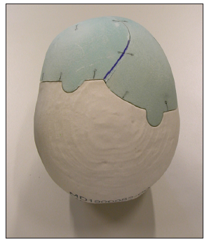
Figure 1. "Puzzle" technique. The perimeters of cranioplasty must be characterized by extroflexions to prevent slips and dislocations
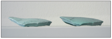
Figure 2. Forehead cranioplasty. The edges have an inclination of 45° to prevent the sinking of the cranioplasty. Male forehead profile on the left; female forehead profile on the right
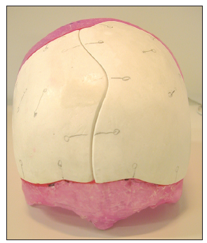
Figure 3. "Italic S" technique. If the cranioplasty involves the use of more pieces faced between them, the contact surfaces must not be linear. This prevents slips and dislocations
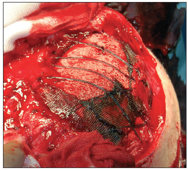
Figure 4. In the pterional area, the anchorage of the temporalis muscle should not be done on the cranioplasty, but must override it, with traction to the sagittal line
Attempts should also be made to prevent the formation of a fluid fistula, which can severely slow or impede cicatrisation and osteomimesis. The main cause of fistulas is adhesion between the dura mater, temporal muscle, and galea.[14] Such scarring adhesions can prolong subsequent surgery times, cause excessive blood loss, and increase the probability of an inadvertent lesion to the dura mater or cerebral cortex due to the difficult techniques required for their dissection.[15] Nevertheless, these events can be averted by placing an inert, nonresorbable membrane, such as a super-thin (0.1 mm) sheet of expanded polytetrafluoroethylene (ePTFE; e.g. PrecludePeritoneal Membrane, W.L. Gore and Associates. Inc.), between the dura and the soft tissues, especially at the site of the temporal muscle.[16]
Extradural and extracranial pooling of fluid and subdural hematoma are less frequent events in cranioplasties. The former can usually be resolved by prompting parenchymal re-expansion (if viable) or by increasing the number of dural suspension points and maintaining subcutaneous drainage for a longer period of time. Adhesion of the scalp to the cranial implant can be promoted by anchoring the latter to the galea fascia using sutures.
The soft tissues overlying the cranioplasty implant can also be subject to ischemia, necrosis, and/or decubitus, and it is thus vital that cutaneous trophism and irrigation is carefully evaluated in the presurgical phase. Moreover, a surgical approach should be planned taking into account not only aesthetic concerns (such as avoiding the incision encroaching below the hairline and using the Simpson technique) but also seeking to avoid damage to the main arterial trunks and temporal muscle.[13,17] In difficult cases featuring a paucity of viable soft tissue, cranioplasty implant fitting could necessitate the use of cutaneous expanders. Another useful surgical aid for improving cutaneous trophism is dermal matrix (INTEGRA Dermal Regeneration Template Single Layer film) [Figure 5].[18] Such matrices promote mesenchymal histoinduction and histoconduction, serving to guide the formation of normal dermal tissue. The collagen and glucosaminoglycans of these matrices provide structural support for the infiltrating fibroblasts, macrophages, lymphocytes, and capillaries that form the neurovascular network. In covering the implant, these networks favor the development of better blood irrigation, important not only for cutaneous tropism but also for the invasion of the porous HA of the cranial implant by the organic bone matrix, promoting osteoconduction and osteointegration of the prosthesis. The scalp is not only necessary for implant coverage but it also supplies nutrients and immune system components. Together with the dura, it also aids in the osteomimesis process of the cranioplasty implant.
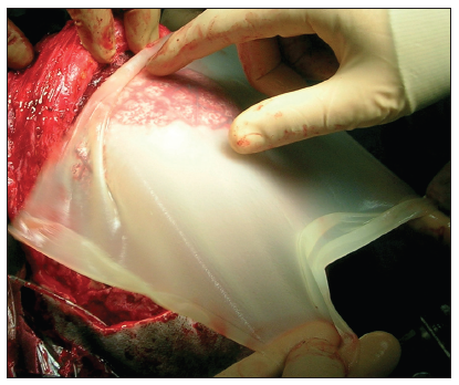
Figure 5. The trophism of the skin overlying the cranioplasty is important for the osteomimesis and for the prevention of infections. The trophism of the skin may improve by using dermal matrix placed between cranioplasty and subcutaneous tissue
Indeed, another possible cause of HA cranial implant failure is lack of osteomimesis. If there is poor contiguity between the implant and the skull margin, osteoblast migration is compromised. To avoid this and to ensure the accurate design of the implant (which must fit perfectly along the entire border of its cranial housing), the surgeon must take certain factors into account during the surgery itself. In particular, the skull defect borders must be cleared completely of any scarring or inflammatory matrix, the dura on the border of the internal plate must be delaminated, and the craniectomy border drilled delicately. In addition, no material should be placed between the bone and implant, with the exception of HA granules or calcium phosphate paste [Figure 6]. Indeed, it has been demonstrated that more osteointegration occurs on a rough surface.[19] A prime concern of the surgeon, however, should be that the continuum is controlled and that the tissue exposed to drilling is adequately cooled.
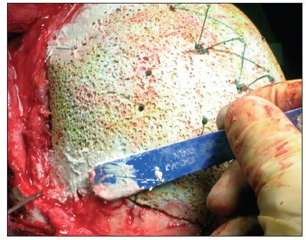
Figure 6. The solutions of continuous between cranioplasty and be must be filled with moldable pastes of calcium phosphate. This promotes osteointegration and increases the primary resistance of the cranioplasty thanks to optimal bone perimeter support (curb)
In fact, the threshold for damage to osteocytes is as low as 47 °C.[20] That being said, the limited clinical success of osteomimesis could also be explained by a lack of vascularization, which is affected by the tropism of the overlying skin, a critical process during bone growth and repair.[21]
In general, it appears that the majority of adverse events in cranioplasties are ascribable to human error, on the part of the manufacturer or the surgeon. Indeed, poor design or lack of adequate preparation is responsible for almost all custom-made HA cranioplasty implant failures. For this reason, a continuous exchange of information among surgeons and implant manufacturing technicians is essential, and should go some way to ensuring the continued success of this procedure.
Financial support and sponsorship
Nil.
Conficts of interest
There are no conflicts of interest.
REFERENCES
1. Nataloni A. The biomimetism to medical device quality. Riv Med 2005;11:133-4.
2. Guidelines on a Medical Devices Vigilance System. MEDDEV 2.12-1 rev. 6 December 2009, European Commission; DG Enterprise and Industry. Available from: http://www.ec.europa.eu/health/medical-devices/files/meddev/2_12_1-rev_6-12-2009_en.pdf. [Last cited on 2014 Dec 5].
3. Eufinger H, Wehmöller M. Individual prefabricated titanium implants in reconstructive craniofacial surgery: clinical and technical aspects of the first 22 cases. Plast Reconstr Surg 1998;102:300-8.
4. Joffe J, Harris M, Kahugu F, Nicoll S, Linney A, Richards R. A prospective study of computer-aided design and manufacture of titanium plate for cranioplastyand its clinical outcome. Br J Neurosurg 1999;13:576-80.
5. Agner C, Dujovny M, Park H. Delayed minimally invasive cranioplasty. Minim Invasive Neurosurg 2003;46:186-90.
6. Chiarini L, Figurelli S, Pollastri G, Torcia E, Ferrari F, Albanese M, Nocini PF. Cranioplasty using acrylic material: a new technical procedure. J Craniomaxillofac Surg 2004;32:5-9.
7. Lee SC, Wu CT, Lee ST, Chen PJ. Cranioplasty using polymethyl methacrylate prostheses. J Clin Neurosci 2009;16:56-63.
8. White JC. Late complications following cranioplasty with alloplastic plates. Ann Surg 1948;128:743-54.
9. Gürbüz MS, Celik O, Berkman MZ. Infection of cranioplasty seen twenty years later. J Korean Neurosurg Soc 2012;52:498-500.
10. De Bonis P, Frassanito P, Mangiola A, Nucci CG, Anile C, Pompucci A. Cranial repair: how complicated is filling a "hole"? J Neurotrauma 2012;29:1071-6.
11. Zanotti B, Cramaro A. Notes of surgical procedure. Riv Med 1995;11:153-60.
12. Johnson PJ, Robbins DL, Lydiatt WM, Moore GF. Salvage of an infected hydroxyapatite cement cranioplasty with preservation of the implant material. Otolaryngol Head Neck Surg 2000;123:515-7.
13. Zanotti B, Verlicchi A, Robiony M, Parodi PC. Surgical calvarial demolition and reconstruction: procedure, implants and results. Top in Med 2010;16:1-19.
14. Kim H, Sung SO, Kim SJ, Kim SR, Park IS, Jo KW. Analysis of the factors affecting graft infection after cranioplasty. Acta Neurochir (Wien) 2013;155:2171-6.
15. Chun HJ, Yi HJ. Efficacy and safety of early cranioplasty, at least within 1 month. J Craniofac Surg 2011;22:203-7.
16. Zanotti B, Ius T. Surgery: useful to know. Riv Med 2005;11:185-7.
17. Zanotti B, Verlicchi A. Craniectomia decompressiva. In: Zanotti B, Verlicchi A, Parodi PC, editors. Cranioplastica Terapeutica. Trento: New MAGAZINE edizioni; 2013. pp. 37-46.
18. Cordaro ER, Calabrese S, Faini GP, Zanotti B, Verlicchi A, Parodi PC. Method to thicken the scalp in calvarian reconstruction. J Craniofac Surg 2011;22:598-601.
19. Albrektsson T, Wennerberg A. Oral implant surfaces: part 1 - review focusing on topographic and chemical properties of different surfaces and in vivo responses to them. Int J Prosthodont 2004;17:536-43.
20. Dolan EB, Haugh MG, Tallon D, Casey C, McNamara LM. Heat-shock-induced cellular responses to temperature elevations occurring during orthopaedic cutting. J R Soc Interface 2012;9:3503-13.
Cite This Article
Export citation file: BibTeX | RIS
OAE Style
Zanotti B, Verlicchi A, Stefini R, Salgarelli AC, Zingaretti N, Parodi PC, Matteo C, Robiony M. Surgical pitfalls with custom-made porous hydroxyapatite cranial implants. Plast Aesthet Res 2015;2:7-11. http://dx.doi.org/10.4103/2347-9264.149364
AMA Style
Zanotti B, Verlicchi A, Stefini R, Salgarelli AC, Zingaretti N, Parodi PC, Matteo C, Robiony M. Surgical pitfalls with custom-made porous hydroxyapatite cranial implants. Plastic and Aesthetic Research. 2015; 2: 7-11. http://dx.doi.org/10.4103/2347-9264.149364
Chicago/Turabian Style
Zanotti, Bruno, Angela Verlicchi, Roberto Stefini, Attilio Carlo Salgarelli, Nicola Zingaretti, Pier Camillo Parodi, Casadei Matteo, Massimo Robiony. 2015. "Surgical pitfalls with custom-made porous hydroxyapatite cranial implants" Plastic and Aesthetic Research. 2: 7-11. http://dx.doi.org/10.4103/2347-9264.149364
ACS Style
Zanotti, B.; Verlicchi A.; Stefini R.; Salgarelli AC.; Zingaretti N.; Parodi PC.; Matteo C.; Robiony M. Surgical pitfalls with custom-made porous hydroxyapatite cranial implants. Plast. Aesthet. Res. 2015, 2, 7-11. http://dx.doi.org/10.4103/2347-9264.149364
About This Article
Copyright

Data & Comments
Data


 Cite This Article 8 clicks

 Like This Article 0
likes
Like This Article 0
likes


Comments
Comments must be written in English. Spam, offensive content, impersonation, and private information will not be permitted. If any comment is reported and identified as inappropriate content by OAE staff, the comment will be removed without notice. If you have any queries or need any help, please contact us at support@oaepublish.com.