
Efficacy of targeted muscle reinnervation for treating and preventing postamputation pain - a systematic review

Abstract
Aim: Targeted muscle reinnervation (TMR) is a procedure pioneered to improve control of myoelectric prostheses and was fortuitously found to improve postamputation pain by transferring residual nerve ends from an amputated limb to reinnervate motor nerve units in denervated muscles. This study sought to perform a systematic review of the literature regarding the postamputation pain-related outcomes following TMR.
Methods: PubMed database was queried using the key term “targeted muscle reinnervation”. Articles were chosen based on the following criteria: (1) clinical studies on TMR; (2) greater than one subject; (3) studies were case-controls, comparative cohort analyses, controlled trials, or randomized controlled trials; and (4) studies included one or more outcomes of interest: prosthetic use and functionality, improvement or persistence of pain, indications, complications, donor nerves, and technical aspects of TMR.
Results: Overall, 9 studies including 101 upper extremity and 252 lower extremity nerve transfers were analyzed, with nerve transfer type, amputation location, and specific neurotizations reported. Four studies assessed the efficacy of TMR in addressing phantom limb pain (PLP) and residual limb pain (RLP), with 3 out of 4 studies reporting significant improvements in PROMIS (Patient Reported Outcome Measurement Information System) scores in TMR subjects compared to controls. Five additional studies did not analyze PROMIS scores but reported subjective improvements in pain outcomes.
Conclusion: Included studies demonstrated TMR had lower maximal pain and pain intensity, behavior and interference compared to the standard of care. Secondary TMR used to treat patients with established painful neuromas also reported improvement in pain compared to baseline.
Keywords
INTRODUCTION
Targeted muscle reinnervation (TMR) is a nerve transfer procedure originally pioneered to improve the myoelectric control of upper limb prostheses by transferring residual mixed or sensory nerve ends from an amputated limb to reinnervate target motor nerve units in denervated muscles[1-3]. Once surgically relocated, the fascicles of the transferred nerve will grow into the recipient muscle motor end plates[4]. This procedure allows the creation of additional signals that can be used to enhance myoelectric prosthetic control and optimize function[5]. In addition to more intuitive control of myoelectric prostheses, patients who underwent TMR reported better outcomes with common amputation complications, particularly neuroma pain. As a result, TMR has recently been adopted as an effective strategy for the management and prevention of postamputation pain, including neuroma pain, phantom limb pain (PLP), and residual limb pain (RLP)[6,7].
There are multiple distinct types of pain that a patient may experience postamputation. PLP is defined as the perception of burning, tingling, discomfort, or electrical shooting pain in the missing portion of the limb[6,8,9]. This pain may be localized to just one region of the missing limb or may extend over the entire missing area. PLP typically occurs within the first 6 months postamputation, although its prevalence several years after surgery has been reported to be as high as 85%[10-12]. RLP, also known as “stump” pain, is localized to the portion of the limb remaining after the amputation. RLP is typically described as a sharp, electrical, burning, or “skin-sensitive” pain that may be localized superficially at an incision or deep in the residual limb. It can also encompass the entirety of the residual limb. The reported incidence of stump pain can be as high as 74% and, like PLP, may persist for years after initial development[10-13]. RLP may also be driven by terminal symptomatic neuromas that become irritated by pressure, light touch, and hot or cold temperatures[8,9]. Although neuromas may be a cause of RLP, neuroma pain is distinct from RLP and occurs due to uncoordinated attempts of nerve fibers to regenerate, resulting in disorganized axons encased within scar tissue at the site of nerve transection or injury. They are responsible for much of the RLP experienced postamputation and may be difficult to treat with high recurrence rates[1].
Despite the increasing use of TMR for improvement of postamputation pain, there are few studies comparing the functional outcomes of patients who underwent TMR procedures primarily for this purpose. This study sought to perform a systematic review of the literature regarding the outcomes of postamputation pain in patients who have undergone TMR procedures, including RLP, PLP, and neuroma pain.
METHODS
This study was conducted in accordance with the Preferred Reporting Items for Systematic Reviews and Meta-Analyses (PRISMA) guidelines[14]. The PubMed database was queried for articles published in English as the primary language in May 2021. A Boolean operator with the key term “targeted muscle reinnervation” was employed to conduct the search. 588 articles were found and sorted using the “Best Match” criteria. For each relevant article, additional articles were searched for using the “Similar Articles” section as part of the systematic screening process to identify articles that may have been missed by the original search query. Articles were chosen based on the following criteria: (1) studies were clinical studies on TMR; (2) studies included greater than one subject; (3) studies were either case-controls, comparative cohort analyses, controlled trials, or randomized controlled trials; and (4) studies included one or more outcomes of interest. Outcomes of interest included: prosthetic use and functionality, improvement or persistence of pain, indications, complications, donor nerves, and technical aspects of TMR. Case reports and letters to the editor were excluded. There were no restrictions on the year of publication. After the articles identified through the original query through the PubMed database were screened, the full-text articles were assessed for eligibility and inclusion in qualitative synthesis. Twenty-seven additional articles were surveyed from “Similar Articles”; of the 615 total studies, 9 studies met the final inclusion criteria [Figure 1]. In accordance with PRISMA guidelines, 2 reviewers independently assessed the quality and methodology of each study[14].
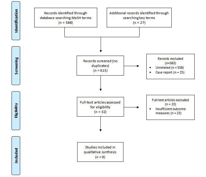
Figure 1. PRISMA flow diagram of included studies.
RESULTS
As part of the systematic review, nerve transfer type, amputation location, and specific neurotizations were reported [Table 1]. Overall, 101 upper extremity nerve transfers were analyzed (with 11 of these specifically reported as primary TMR for the upper extremity and 8 reported as secondary TMR for the upper extremity). Specified amputation locations included trans-radial (19), trans-humeral (38), shoulder disarticulation/glenohumeral (32), above-elbow (8), below-elbow (5), elbow disarticulation (1), and CMC joint (1) amputations. Neurotizations for the upper extremity primarily involved the ulnar, median, radial, and musculocutaneous nerves, although additional nerves (including the medial cord, lateral cord, posterior cord, radial, intercostal, and intercostal brachial cutaneous) were also involved in nerve transfer. A variety of muscle targets for the upper extremity were identified, and selected based on amputation level, patient-specific anatomy, zone of injury, mechanism of injury, and nerve length[7,8,15-21].
Included studies in systematic review
| First author, year | Amputation location (n), Primary/Secondary TMR | Amputation location (n) | Neurotizations |
| Alexander, 2019[6] | Upper extremity (9), Primary | Trans-radial (1) Trans‐humeral (4) Shoulder disarticulations (4) | Not specified |
| Lower extremity (22), Primary | Below-knee (7) Above-knee (14) Hip disarticulation (1) | Not specified | |
| Dumanian, 2019[7] | Upper extremity (4), Secondary | Above-elbow (3) Below-elbow (1) | ● Not specified |
| Lower extremity (26), Secondary) | Above-knee (10) Below-knee (16) | ● Not specified | |
| Janes, 2020[15] | Lower extremity (17) | Trans-tibial (7), Secondary Trans-femoral (10), Primary | ● Saphenous nerve → Medial soleus muscle, medial gastrocnemius biceps femoris, vastus medialis, gracilis |
| Kubiak, 2019[16] | Upper extremity (6), Primary | Trans-radial (3) Trans-humeral (2) Glenohumeral (1) | ● Median cord, lateral cord, posterior cord, musculocutaneous, ulnar, median, radial, SBRN, intercostal brachial cutaneous nerves |
| Lower extremity (46), Primary | Trans-tibial (37) Trans-femoral (9) | ● Sciatic, femoral, tibial, common peroneal, deep peroneal, superficial peroneal, saphenous, sural | |
| Morgan, 2016[17] | Upper extremity (5) | Trans-radial (3), Primary; (2) Secondary | ● Median nerve → FDS, FDP ● Ulnar nerve → FCU, FPL ● SBRN → Extensor carpi radialis, FDS |
| Pet, 2014[18] | Upper extremity (11), Primary | Elbow disarticulation (1) Long trans-humeral (3) Short trans-humeral (4) Above-elbow (1) Shoulder disarticulation (2) | ● Median nerve → Medial biceps, biceps, FDS, pectoralis major, lateral biceps, upper pectoralis major ● Radial nerve → Teres minor lateral triceps, medial triceps, serratus anterior, lateral triceps, brachialis, triceps, latissimus dorsi, lateral FDS ● Ulnar nerve → Medial triceps, serratus, lower pectoralis major, teres minor, triceps, posterior triceps, pectoralis minor, triceps, FDS ● Musculocutaneous nerve → Clavicular head of pectoralis major, medial biceps, lateral biceps, biceps |
| Upper extremity (8), Secondary | CMC joint (1) Trans-radial (3) Trans-humeral (3) Shoulder disarticulation (1) | ● Median nerve → Clavicular head of pectoralis major, medial biceps, FDS, FDP ● Radial nerve → Lateral triceps, medial triceps, brachialis, FDS ● Ulnar nerve → Medial triceps, posterior triceps, FDS | |
| Lower extremity (1), Primary | Knee disarticulation (1) | ● Tibial nerve → Medial hamstring, hamstring ● Peroneal nerve → Lateral hamstring | |
| Lower extremity (15), Secondary | Above-knee (8) Below-knee (7) | ● Sciatic nerve → Lateral hamstring, medial hamstring ● Tibial nerve → Medial hamstring, hamstring ● Peroneal nerve → Lateral hamstring | |
| Souza, 2014[19] | Upper extremity (26), Secondary | Trans-humeral (16) | ● Median nerve → Biceps brachii (short head) ● Ulnar nerve → Brachialis ● Radial nerve → Triceps brachii (lateral head) |
| Shoulder disarticulation (10) | ● Musculocutaneous nerve → Pectoralis major (clavicular head) ● Median nerve → Pectoralis major (split sternal head) ● Ulnar nerve → Pectoralis major (split sternal head), pectoralis minor, latissimus dorsi, serratus anterior ● Radial nerve → Latissimus dorsi, serratus anterior, pectoralis major (split sternal head) | ||
| Valerio, 2019[20] | Upper extremity (15), Primary | Above-elbow (4) Below-elbow (4) Shoulder disarticulation (7) | Not Specified |
| Lower extremity (36), Primary | Above/through knee (18) Below-knee (18) | Not Specified | |
| Obrien, 2021[21] | Upper extremity (16), Primary | Trans-radial (5) | ● Median nerve → FDS or FDP ● Ulnar nerve → FCU or FPL ● SBRN → Lateral head of triceps ● MABC nerve → Brachioradialis, FDP, ECRL LABC nerve → ECRL, ECRB |
Transhumeral (5) | ● Median nerve → Short head of biceps ● Ulnar nerve → Brachialis ● Radial nerve → Lateral head of triceps ● Medial antebrachial cutaneous nerve → Brachialis ● Musculocutaneous nerve → Short head, long head of biceps | ||
Shoulder disarticulation (6) | ● Musculocutaneous nerve → Clavicular head of pectoralis major ● Median nerve → Sternal head of pectoralis major ● Ulnar nerve → Sternal head of pectoralis major ● Radial nerve → Tibial nerve, latissimus dorsi |
For lower extremity amputations, 252 were reported, with specific amputation sites including below-knee (48), above-knee (50), hip disarticulation (1), trans-tibial (82), trans-femoral (15), and knee disarticulation (1). Neurotizations for the lower extremity primarily involved the tibial, saphenous, sciatic, and peroneal nerves, although additional nerves including the posterior femoral cutaneous nerve, femoral, and sural nerve (among others) were used as well[7,8,15-20].
A total of four studies assessed the possible benefits of TMR in PLP and RLP via PROMIS (Patient Reported Outcome Measurement Information System) [Table 2][7,8,20,21]. PROMIS is a self-reporting tool to capture respondents’ perception of pain through its impact on multiple components of daily life including physical, social, and emotional pillars. It utilizes three aspects-intensity, behavior, and interference. Intensity is represented by standardized pain rating scales (verbal, numerical, visual analog). Pain Interference applies a numerical rating score for degree of impedance in professional, familial, emotional, and recreational life. Pain behavior applies a similar numerical rating in the context of how one specifically acts or reacts through observable displays or phonation. All these studies, with the exception of Dumanian et al., reported significant improvements in PROMIS parameters in TMR subjects compared to controls[7]. PROMIS analysis was also performed for the subcategory of worst pain, as outlined in Table 3.
PROMIS analysis - worst pain
| First author, year | Worst pain at baseline | Worst pain at 1 year | Change from baseline | Worst pain at last follow-up | Change from baseline | |
| Dumanian, 2019[7] | PLP TMR | 5.8 (SD 3.2) | 2.6 (2.2) | 3.2 (2.9) | 2.3 (2.3) | 3.5 (3.1) |
| Standard | 3.9 (SD 2.7) | 4.1 (3.0) | -0.2 (4.9) | 4.4 (3.3) | -0.5 (5.3) | |
| RLP TMR | 6.6 (2.0) | 3.7 (2.0) | 2.9 (2.2) | 3.6 (2.1 | 3.0 (2.1) | |
| Standard | 6.9 (2.5) | 6.0 (2.5) | 0.9 (3.3) | 5.7 (3.0) | 1.2 (3.5) |
PROMIS analysis - pain intensity, behavior & interference
| Pain intensity | Pain behavior | Pain interference | Follow-up | ||
| Alexander, 2019[6] | PLP (mean differences) | 5.855 (95%CI 1.159, 10.55; P = .015) | 5.896 (95%CI 0.492, 11.30; P = .033) | 7.435 (95%CI 1.797, 13.07; P = .011) | > 1 year |
| RLP (mean differences) | 5.477 (95%CI 0.528, 10.42; P = .031) | 6.195 (95%CI 0.705, 11.69; P = .028) | 6.816 (95%CI 1.438, 12.2; P = .014) | > 1 year | |
| Dumanian, 2019[7] | PLP (mean differences) | 11.7 (-0.3, 23.7) | 1.1 (-8.3, 10.5) | 4.7 (-5.0, 14.3) | At 1 year |
| 9.3 (-1.4, 20.0) | 4.3 (-4.7, 13.2) | 4.7 (-5.6, 15.3) | At last follow-up | ||
| RLP (mean differences) | 5.8 (-0.9, 12.4) | -0.5 (-7.2, 6.1) | -0.9 (-8.5, 6.7) | At 1 year | |
| 5.8 (-0.3, 11.2) | -0.7 (-7.5, 6.1) | 0.5 (-7.0, 8.1) | At last follow-up | ||
| Valerio, 2019[20] | PLP (median t-score) | TMR 36.3 vs. control 48.3 | TMR 50.1 vs. control 56.6 | TMR 40.7 vs. control 55.8 | |
| RLP (median t-score) | TMR 30.7 vs. control 46.8 | TMR 36.7 vs. control 57.3 | TMR 40.7 vs. control 57.3 | Median 330 days (TMR group) | |
| O’Brien, 2021[21] | PLP (median t-score) | 33.5 vs. control 46.8 P = < .05 | 50.1 vs. control 53.1 P = < .05 | 40.7 vs. control 50 P = < .05 | Average 23.1 months (for TMR group) |
| RLP (median t-score) | 33.5 vs. control 46.8 P = < .05 | 36.7 vs. control 53.1 P = < .05 | 40.7 vs. control 48.2 P = .146 |
Several studies included in the systematic analysis did not analyze PROMIS scores, however, still reported patient subjective improvements in pain outcomes including neuroma pain [Table 4]. Janes et al reported that of the 10 patients who underwent TMR for chronic neuroma pain, 7 patients (those not lost to follow-up) were seen an average of 4 months postoperatively, with 2 reporting reduced neuroma pain and 5 reporting complete resolution of pain[15]. Of the 7 patients who underwent acute TMR at the time of amputation for prevention of neuroma pain and postamputation pain, the 3 patients not lost to follow-up (seen on average 6.67 months postoperatively) denied development of neuroma pain.
Subjective patient outcomes
| First author, year | Type of study | Nerve transfer | Outcome |
| Janes, 2020[15] | Case series | Lower extremity (17) ● Primary treatment for neuroma pain (10) ● Secondary treatment (7) | Primary treatment 7 total follow-up patients: 5 reported resolution of symptoms, 2 reported improvement in pain Secondary treatment: all denied development of neuroma pain |
| Kubiak, 2019[16] | Retrospective cohort | Primary treatment Upper extremity (10) Lower extremity (80) | 0 patients in treatment group developed symptomatic neuroma vs. 13.3% of control 51.1% of TMR patients developed PLP vs. 91.1% |
| Morgan, 2016[17] | Case series | Upper extremity (5) ● Primary treatment for neuroma pain (3) ● Secondary treatment (2) | All patients reported improvements in pain symptoms |
| Pet, 2014[18] | Retrospective review | Upper extremity (19) ● Primary treatment (11) ● Secondary treatment (8) Lower extremity (16) ● Primary treatment (1) ● Secondary treatment (15) | 92% of primary TMR treatment neuroma free. 50% developed PLP 87% of secondary TMR treatment neuroma free. Equivocal findings regarding PLP |
| Souza, 2014[19] | Retrospective Review | Secondary treatment Upper extremity (26) | 93% of patients with existing neuroma pain experienced resolution of symptoms |
Additional studies reported outcomes for neuroma pain[15-19]. Kubiak et al reported postoperative outcomes in a total of 90 patients, with 45 of these patients acting as controls and 45 undergoing TMR[16]. 6 control patients (13.3%) developed symptomatic neuromas in the postoperative period, compared with 0 patients in the TMR group (P = 0.026). 23 TMR patients (51.1%) reported the development of PLP, compared with 41 control patients (91.1%; P < 0.0001)[16]. Likewise, Morgan et al reported that among 3 patients undergoing revision amputation with TMR for treatment of painful neuromas and 2 patients undergoing elective amputation with concurrent TMR, all 5 patients reported improvement in pain[17]. Although all 5 reported improvements in pain, only 4 were able to use a prosthesis following the procedure. Souza et al. reported that of 15 patients presenting with preexisting neuroma pain, 14 experienced complete resolution of pain after TMR, with 1 patient having improvement of neuroma pain. No patients reported new-onset neuroma pain following the TMR procedure[19]. Pet 2014 analyzed 12 patients undergoing primary TMR for neuroma prevention and 23 patients with established neuromas who underwent neuroma excision with secondary TMR and reported that at follow-up, 11 of 12 patients (92%) after primary TMR and 20 of 23 patients (87%) after secondary TMR were free of palpation-induced neuroma pain. Of the cohort undergoing primary TMR, 6 out of 12 patients did develop PLP. For those undergoing secondary TMR, PLP was present in 8 patients before secondary TMR and in 8 patients afterward, showing persistent PLP in 7 patients with new onset of phantom pain in 1 patient, and resolution of preoperative phantom pain in 1 patient[18].
DISCUSSION
This systematic review of currently available literature supports the use of TMR to minimize PLP and residual limb pain (RLP) after upper and lower extremity amputation. Included studies demonstrated patients undergoing TMR had lower maximal pain and pain intensity, behavior and interference compared to the standard of care of burying the cut nerve in muscle. Secondary TMR used to treat patients with established painful neuromas also reported an improvement in their pain compared to their preoperative baseline. With encouraging outcomes having been reported throughout multiple randomized controlled trials, TMR is emerging as a leading surgical technique for pain prevention in patients undergoing major limb amputations and pain management in patients with preexisting amputations.
TMR has proven a useful tool in accentuating myoelectric potential in prosthesis as well as improving pain outcomes, specifically in regard to PLP and RLP[22,23]. The overall morbidity of PLP and RLP in amputees has been reported in several studies with rates as high as 67% and 25%, respectively[24]. This systematic review serves to specifically identify outcome parameters related to these debilitating sequelae of amputation, inclusive of diverse etiologies and timing related to index and subsequent procedures. While studies measure TMR outcomes in a variety of manners, our review underscores PROMIS for its versatility and inclination to the multifaceted nature of PLP and RLP. Developed by the NIH, The PROMIS explores person-centered agency using physical, mental, and social facets of one’s health. Researchers involved in TMR have found this tool particularly relevant in capturing holistically the devastating effects of PLP and RLP on physical and emotional well-being.
Our investigation identified four studies meeting inclusion criteria assessing PLP/RLP; all of which showed improvements in outcome parameters. Only one study, Dumanian et al, with limited patient enrollment
The remaining studies were retrospective cohort studies and demonstrated statistical significance across PROMIS components including pain intensity, pain behavior, and pain interference. Alexander et al. uniquely studied amputations related to oncologic treatment with concurrent TMR and incorporated follow-up to 1 year[6]. This study also had limited total patients (31 TMR). Although TMR was done at the time of amputation, only 16 patients underwent TMR at index surgery. The remaining underwent secondary amputation related to recurrence or infection. These patients were also affected by neoadjuvant/adjuvant chemotherapy and radiation.
Valerio et al. focused on the general amputee population with larger patient numbers totaling 438 subjects, 51 of which had TMR performed at index procedure[20]. Subjects represented diverse ages, levels of amputation, and indications for amputation. The aggregate data perhaps favors a more generalized representation, emphasizing marked improvements in PROMIS reporting across intensity, behavior, and interference. The follow-up ranged 3 months to 5.3 years (> 1 year 64.7%). One limitation, however, is that the follow-up survey of the non-TMR cohort was noted to be longer after surgery, given the retrospective nature. Long-term data are needed to determine if TMR results are consistent over time without recurrence of functional limitation. This likely introduced respondent reporting biases.
The latest 2021 study by O’Brien et al was also a retrospective cohort study, which included 16 patients who underwent TMR at index amputation compared to 55 controls. 62% of the TMR patients had no PLP versus 24% of controls. Similarly, half of TMR patients were without RLP versus 36% of controls. PROMIS scores across all parameters, with the exception of RLP interference, significantly favored TMR[21].
Although PROMIS scores offer a tremendous metric for assessing the debilitating pillars of RLP and PLP, it is not without limitations. Its design remains predicated on an objective iteration of subjectively assigned values in a presumably standardized manner. Moreover, the processes for all the above-mentioned studies were reliant upon patients’ ability to distinguish PLP from RLP, which at times may be tenuous.
Limitations of this study include the lack of meta-analysis, which was not feasible given the wide variation in data points collected among the different studies. Additionally, the information does not allow for outcome conclusions comparing specific nerve transfers. Generally, target motor nerves in both upper and lower extremity TMR are ideally those which have redundancy in motor function to maintain physiologic continuity. The target nerve should be an expendable nerve preserving another nerve that has similar functions. This is particularly relevant in below knee amputation, where the larger medial gastrocnemius is preserved to provide adequate protective bulk for prosthesis fitting. Despite the statistical limitations and inability to compare transfers across multiple studies, this review supports the use of TMR in the prevention and treatment of RLP and PLP.
This study includes new literature not evaluated in prior reviews. This is critical given the recent increase in the adoption of and research related to this technique[25,26]. This study is also unique due to its strict inclusion criteria of control groups to underscore clinical and patient-reported outcome measures in standard practice. Additionally, our appraisal of the literature emphasized specifically studies that assessed PROMIS, a metric we believe is especially meaningful in capturing the multifaceted nuances of living with postamputation pain syndromes.
This systematic review adds to the literature supporting the efficacious use of TMR, both as a technique to improve postamputation pain compared to previously established standards of care and as a treatment for established postamputation neuromas.
DECLARATIONS
Authors’ contributionsWriting, review and editing: Di Valerio E, Lautenslager L, Vonu P, Mustafa C, Chim H, Satteson E
Original draft and methodology: Di Valerio E, Lautenslager L, Vonu P, Satteson E
Data curation: Di Valerio E, Lautenslager L
Formal analysis: Lautenslager L, Vonu P
Conceptualization: Vonu P, Mustafa C, Chim H, Satteson E
Supervision, project administration, resources: Satteson E
Availability of data and materialsNot applicable.
Financial support and sponsorshipNone.
Conflicts of interestAll authors declared that there are no conflicts of interest.
Ethical approval and consent to participateNot applicable.
Consent for publicationNot applicable.
Copyright© The Author(s) 2022.
REFERENCES
1. Cheesborough JE, Smith LH, Kuiken TA, Dumanian GA. Targeted muscle reinnervation and advanced prosthetic arms. Semin Plast Surg 2015;29:62-72.
2. Chappell AG, Jordan SW, Dumanian GA. Targeted muscle reinnervation for treatment of neuropathic pain. Clin Plast Surg 2020;47:285-93.
3. Hijjawi JB, Kuiken TA, Lipschutz RD, Miller LA, Stubblefield KA, Dumanian GA. Improved myoelectric prosthesis control accomplished using multiple nerve transfers. Plast Reconstr Surg 2006;118:1573-8.
4. Kuiken TA, Dumanian GA, Lipschutz RD, Miller LA, Stubblefield KA. The use of targeted muscle reinnervation for improved myoelectric prosthesis control in a bilateral shoulder disarticulation amputee. Prosthet Orthot Int 2004;28:245-53.
5. Bergmeister KD, Aman M, Muceli S, et al. Peripheral nerve transfers change target muscle structure and function. Sci Adv 2019;5:eaau2956.
6. Alexander JH, Jordan SW, West JM, et al. Targeted muscle reinnervation in oncologic amputees: early experience of a novel institutional protocol. J Surg Oncol 2019;120:348-58.
7. Dumanian GA, Potter BK, Mioton LM, et al. Targeted muscle reinnervation treats neuroma and phantom pain in major limb amputees: a randomized clinical trial. Ann Surg 2019;270:238-46.
9. Buchheit T, Van de Ven T, Hsia HL, et al. Pain phenotypes and associated clinical risk factors following traumatic amputation: results from veterans integrated pain evaluation research (VIPER). Pain Med 2016;17:149-61.
10. Hsu E, Cohen SP. Postamputation pain: epidemiology, mechanisms, and treatment. J Pain Res 2013;6:121-36.
11. Jensen TS, Krebs B, Nielsen J, Rasmussen P. Immediate and long-term phantom limb pain in amputees: Incidence, clinical characteristics and relationship to pre-amputation limb pain. Pain 1985;21:267-78.
13. Ehde DM, Czerniecki JM, Smith DG, et al. Chronic phantom sensations, phantom pain, residual limb pain, and other regional pain after lower limb amputation. Arch Phys Med Rehabil 2000;81:1039-44.
14. Moher D, Liberati A, Tetzlaff J, Altman DG. PRISMA Group. Preferred reporting items for systematic reviews and meta-analyses: the PRISMA statement. Ann Intern Med 2009;151:264-9,W64.
15. Janes LE, Fracol ME, Ko JH, Dumanian GA. Management of unreconstructable saphenous nerve injury with targeted muscle reinnervation. Plast Reconstr Surg Glob Open 2020;8:e2383.
16. Kubiak CA, Kemp SWP, Cederna PS, Kung TA. Prophylactic regenerative peripheral nerve interfaces to prevent postamputation pain. Plast Reconstr Surg 2019;144:421e-30e.
17. Morgan EN, Kyle Potter B, Souza JM, Tintle SM, Nanos GP 3rd. Targeted muscle reinnervation for transradial amputation: description of operative technique. Tech Hand Up Extrem Surg 2016;20:166-71.
18. Pet MA, Ko JH, Friedly JL, Mourad PD, Smith DG. Does targeted nerve implantation reduce neuroma pain in amputees? Clin Orthop Relat Res 2014;472:2991-3001.
19. Souza JM, Cheesborough JE, Ko JH, Cho MS, Kuiken TA, Dumanian GA. Targeted muscle reinnervation: a novel approach to postamputation neuroma pain. Clin Orthop Relat Res 2014;472:2984-90.
20. Valerio IL, Dumanian GA, Jordan SW, et al. Preemptive treatment of phantom and residual limb pain with targeted muscle reinnervation at the time of major limb amputation. J Am Coll Surg 2019;228:217-26.
21. O’Brien AL, Jordan SW, West JM, et al. Targeted muscle reinnervation at the time of upper-extremity amputation for the treatment of pain severity and symptoms. J Hand Surg Am 2021;46:72.e1-72.e10.
22. Lanier ST, Jordan SW, Ko JH, Dumanian GA. Targeted muscle reinnervation as a solution for nerve pain. Plast Reconstr Surg 2020;146:651e-63e.
23. Peters BR, Russo SA, West JM, Moore AM, Schulz SA. Targeted muscle reinnervation for the management of pain in the setting of major limb amputation. SAGE Open Med 2020;8:2050312120959180.
24. Frantz TL, Everhart JS, West JM, Ly TV, Phieffer LS, Valerio IL. Targeted muscle reinnervation at the time of major limb amputation in traumatic amputees: early experience of an effective treatment strategy to improve pain. JB JS Open Access 2020;5:e0067.
25. McNamara CT, Iorio ML. Targeted muscle reinnervation: outcomes in treating chronic pain secondary to extremity amputation and phantom limb syndrome. J Reconstr Microsurg 2020;36:235-40.
Cite This Article
Export citation file: BibTeX | RIS
OAE Style
Di Valerio E, Lautenslager L, Vonu P, Mustafa C, Chim H, Satteson E. Efficacy of targeted muscle reinnervation for treating and preventing postamputation pain - a systematic review. Plast Aesthet Res 2022;9:62. http://dx.doi.org/10.20517/2347-9264.2022.50
AMA Style
Di Valerio E, Lautenslager L, Vonu P, Mustafa C, Chim H, Satteson E. Efficacy of targeted muscle reinnervation for treating and preventing postamputation pain - a systematic review. Plastic and Aesthetic Research. 2022; 9: 62. http://dx.doi.org/10.20517/2347-9264.2022.50
Chicago/Turabian Style
Di Valerio, Elizabeth, Lauren Lautenslager, Peter Vonu, Chopan Mustafa, Harvey Chim, Ellen Satteson. 2022. "Efficacy of targeted muscle reinnervation for treating and preventing postamputation pain - a systematic review" Plastic and Aesthetic Research. 9: 62. http://dx.doi.org/10.20517/2347-9264.2022.50
ACS Style
Di Valerio, E.; Lautenslager L.; Vonu P.; Mustafa C.; Chim H.; Satteson E. Efficacy of targeted muscle reinnervation for treating and preventing postamputation pain - a systematic review. Plast. Aesthet. Res. 2022, 9, 62. http://dx.doi.org/10.20517/2347-9264.2022.50
About This Article
Special Issue
Copyright

Data & Comments
Data


 Cite This Article 8 clicks

 Like This Article 30
likes
Like This Article 30
likes


Comments
Comments must be written in English. Spam, offensive content, impersonation, and private information will not be permitted. If any comment is reported and identified as inappropriate content by OAE staff, the comment will be removed without notice. If you have any queries or need any help, please contact us at support@oaepublish.com.