
Lower extremity amputation: the emerging role of targeted muscle reinnervation (TMR) and regenerative peripheral nerve interface (RPNI)

Abstract
Lower extremity amputation is increasingly prevalent in the United States, with growing numbers of patients suffering from diabetes and peripheral vascular disease. Amputation has significant functional sequelae as more than half of patients are unable to ambulate at one year postoperatively. Improving mobility and decreasing chronic post-amputation pain can significantly improve the quality of life for these patients and reduce the cost burden on the healthcare system. Plastic and reconstructive surgery has been at the forefront of “reconstructive amputation”, in which nerve pedicles can be surgically guided to decrease painful neuroma formation as well as provide targets for myoelectric prosthesis use. We herein review post-amputation outcomes, epidemiology of chronic, post-amputation pain, and current treatments, including total muscle reinnervation and regenerative peripheral nerve interface, which are at the forefront of multidisciplinary treatment of lower extremity amputees.
Keywords
INTRODUCTION
Extremity amputation is one of the oldest procedures known to man, with archeological records suggesting purposeful amputations as far back as 45,000 years since Neolithic times[1]. The word “amputation” can trace its origin to the Latin term “amputatio” meaning “to cut around”[1]. Amputation continues to be prevalent in the present day, with approximately 1.6 million people with major limb amputations in the United States in 2005 and 185,000 major limb amputations every year[2,3]. By 2050, the prevalence is estimated to be as large as 3.6 million, given that the most common causes for amputation, especially in the lower extremity, are diabetes and peripheral vascular disease, which continue to uptrend[1,3]. Nontraumatic lower extremity amputation is estimated at only 3/10,000 among the healthy population in 2010, but as high as 28.4-46.2/10,000 among diabetics[4]. Unsurprisingly, significant healthcare costs are associated with these patients. The lifetime all-cause direct cost for lower extremity amputation was estimated to be $35.8 billion for the country[4]. No appropriate literature on the indirect or total cost of amputation currently exists, but it is estimated to be significant as studies have found that after a major lower extremity amputation, up to 53.9% of patients were still nonambulatory at 6-month follow-up after the operation[4]. Another retrospective chart review of 206 patients who underwent major lower extremity amputation demonstrated a one-year postamputation ambulatory rate of 46.1%[5]. Furthermore, nonambulatory rates were higher in those with higher BMI, which portend a poor future as the United States population suffers from increasing rates of obesity[5]. Lower extremity amputation is therefore not only a serious problem for individual patients, but a large - and only increasing - burden on the United States healthcare system. We herein aim to discuss post-amputation outcomes, epidemiology of chronic, post-amputation pain, and current treatments including total muscle reinnervation (TMR) and regenerative peripheral nerve interface (RPNI) at the forefront of multidisciplinary treatment of lower extremity amputees.
Outcomes of amputation
Functional equivalence of lower extremity amputation compared to limb salvage was demonstrated in the seminal Lower Extremity Amputation Prevention (LEAP) study[6]. This was a multicenter, prospective, observational study of 569 severe lower extremity injuries who received either amputation or reconstruction[6]. After two years of follow-up, the functional outcome of amputation and reconstruction as measured by the Sickness Impact Profile was not significantly different[6]. Rather, predictors of poor outcome were more correlated with intrinsic patient factors such as education level, ethnicity, insurance status, and social support[6]. Amputation vs. salvage has been a difficult decision tree branching point even for institutions well versed in critical limb injury. Ultimately, Black et al.[7] offer some general consensus, but concluded that many patient-specific factors, as well as injury factors, must be considered prior to decide whether to amputate or salvage. Amputation is therefore not equivalent to failure to salvage, but a viable alternative reconstructive option with likely functional equivalence in the appropriate patient population.
In evaluating functionality after amputation, Suckow et al.[8] performed four focus groups in twenty-six patients who had lower extremity amputations from critical limb ischemia. Quality of life was determined mainly by impaired mobility (65%), pain (60%), progression of disease in the remaining limb (55%), and depression/frustration (54%). Sixty-two percent had multiple prior revascularization procedures. Areas of improvement included peer support (88%), extensive rehab/prosthetist involvement (71%), earlier mention of amputation as a possible outcome (54%), and early discontinuation of narcotics (54%). Physician-controlled factors such as the timing of amputation, informed decision-making, and postamputation support, play an important role[8]. For amputees, the function of their shortened limb correlates closely with prosthetic use[9]. These results have also been corroborated in the LIMB-Q, a patient-reported outcome instrument in which numerous themes were extracted from direct interviews with patients who suffered high-energy lower extremity trauma[10]. Themes such as physical ability, psychological, and prosthesis were important to patients’ daily quality of life[10]. As expected, a long-term study of patients over 20 years after their amputation demonstrated that higher amputation levels were associated with decreased prosthetic use while less intense residual limb pain was associated with greater daily prosthetic use[11]. Therefore, post-amputation pain is not only simply unpleasant, but also directly affects patient function.
POST-AMPUTATION PAIN
Prevalence
Pain significantly impairs the post-amputation patient. In fact, among amputees with chronic pain, it is often not the underlying condition (i.e., amputation of the limb) that primarily limits the individual, but rather the chronic pain they experience[2,9]. Unfortunately, chronic pain is commonplace among amputees. Advances in the measurement of pain, including the validated Patient-Reported Outcomes Measurement Information System (PROMIS) pain interference and pain behavior scores, have been integral in better understanding the significance of pain on patients’ lives[12,13]. PROMIS is a National Institute of Health-funded initiative to develop and validate patient-reported outcomes, including over 300 different measures of physical, mental, and social health typically used for populations with chronic conditions[13]. There are several types of amputation-related pain, including phantom limb pain (PLP), defined as pain in the limb that is no longer present, phantom limb sensation/telescoping, residual limb pain, and back pain[14]. A cross-sectional survey through the Amputee Coalition of America in which 914 amputees were interviewed over the telephone found that 95% of amputees had some daily pain[2]. The most common pain type was phantom limb pain (79.7%), residual limb pain (67.7%), followed by back pain (62.3%)[2]. A systematic review of twelve cross-sectional and three prospective studies of traumatic and atraumatic amputees also reported phantom limb pain incidence as high as 82% at one-year post-amputation and a lifetime prevalence as high as 87%[15]. Even decades after amputation, phantom limb pain, and residual limb pain continue to trouble patients. In a survey of 21 patients who underwent lower limb oncological amputation with a median follow-up duration of 41 years, seventeen reported phantom limb and back pain, fifteen residual limb pain, all with median pain scores five and above on a scale of 1-10[11]. Post-amputation pain is, therefore, a ubiquitous and long-lasting complication of the operation.
Causes of chronic pain
While chronic pain is not yet fully understood, mechanistically, it is due to afferent input from the peripheral nervous system. A randomized, double-blind, placebo-controlled crossover study of lidocaine compared to placebo saline injection in amputees demonstrated expected outcomes of improved pain, especially phantom limb pain, in those who received the lidocaine block[16]. The causes of the pain are thought mainly from chronic nerve compression and neuroma. Chronic nerve compression typically occurs with major nerves at common entrapment sites[17]. Neuromas are classified as neurotmesis or Sunderland 5th degree peripheral nerve injury[18]. When peripheral nerves are injured, the distal end undergoes Wallerian degeneration. The proximal axon is unable to progress to its distal target, and the unorganized fascicular overgrowth results in scarring, thereby forming a neuroma[18]. Psychological factors are also thought to play a major role in both acquisition and maintenance of pain symptoms[19].
Symptomatic neuroma
Neuromas were first described by Abroise Pare in 1634, who treated the symptoms with massage[18]. Neuropathic pain associated with neuroma can be classified into four types: spontaneous pain, pain with pressure over the neuroma, pain on movement of adjacent joints, and dysesthesia or hypersensitivity with light skin touch[20]. Histological features and mechanisms of formation have been explored in several animal studies. A study of Sprague Dawley rat forelimb amputations demonstrated a progression of nerve injury from degenerating axons to axonal spouts to unorganized bundles of axons which grow into muscles and nearby structures and eventually into fibrotic tissue, ultimately forming a neuroma [Figure 1][21]. There are multiple theories on why neuromas cause pain. Proposed mechanisms include repetitive mechanical irritation from tethered scar or compression, myofibroblast proliferation, abnormal accumulation of sodium and potassium channels which cause hyperexcitability, surrounding inflammation that chemically stimulates nearby nociceptors, and release of inflammatory cytokines[18]. The amount of axoplasmic flow, the ratio of fascicles to epineural tissue, and the nutritional status of the peripheral nerve are thought to affect neuroma formation[18].
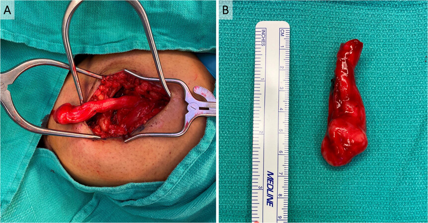
Figure 1. Symptomatic neuroma of sciatic nerve after above-knee amputation (AKA). A 26-year-old man with a history of polyarteritis nodosa who underwent left below-knee amputation as well as right AKA presented with right sciatic nerve symptomatic neuroma approximately 1.5 years after his AKA. The symptomatic neuroma was diagnosed both clinically and radiographically with MRI. (A) In situ sciatic nerve terminal neuroma in his right AKA surgical site. (B) The neuroma was excised, and sciatic nerve cut back to healthy-appearing fascicles. The total resected neuroma and pathologic nerve measured roughly 6 cm long.
Diagnosis of the symptomatic neuroma is based on pain with a scar, altered sensation in the nerve distribution, and positive Tinel’s sign[18]. In cases in which more than one nerve is involved, the picture may be muddled, but the sensory issues should present in a dermatomal pattern[17]. Nerve blocks can be diagnostic and therapeutic in these patients.
Nonsurgical treatment of post-amputation pain
Treatment of post-amputation pain typically begins with non-specialized medical management, including a multimodal pain regimen with nonsteroidal anti-inflammatory drugs, opiates, and non-opiate analgesics like acetaminophen. When pain is not controlled, the patients often get referred to pain specialists where they may receive neuropathic pain medication, including gabapentinoids, antidepressants, or NMDA antagonists like ketamine, dextromethorphan, and memantine[1,22]. These are complemented by physical therapy techniques such as massage or injection therapy, including lidocaine and/or corticosteroid[1]. Regional nerve blocks by pain specialists may serve both diagnostic and therapeutic purposes[17].
Given the significant psychological aspect of post-amputation pain, neurologic and psychologic treatment such as biofeedback and cognitive behavioral therapy are trialed as well[1]. Unique therapies, such as mirror therapy may also be effective. A randomized controlled trial of fifteen amputees underwent mirror vs. control covered mirror or mental visualization therapy[23]. Over four weeks of regular sessions, patients who underwent mirror therapy had a significant decrease in pain scores and daily length of time during which they experienced pain compared to the control group[23]. A systematic review of mirror therapy, motor imagery, and virtual feedback techniques on phantom limb pain after amputation found that while these techniques did seem to reduce pain, the quality of the studies was lacking, and given the limited scientific evidence supporting their effectiveness, no definitive conclusion could be drawn[24]. There has also been active research in neuromodulation techniques. A recent systematic review and meta-analysis of nine randomized controlled trials and five quasi-experimental studies found some hopeful data in transcranial direct current stimulation, although no definitive conclusions were reached yet given the infancy of these techniques for amputees[25].
Multiple disciplines continue to tackle the difficult challenge of post-amputation pain with some exciting preliminary results. However, clinical results of these nonsurgical options continue to be suboptimal, as evidenced by these numerous, diverse, and inconclusive studies. Perioperative management, including assessment of patient and injury risk factors, preoperative consultation of the acute pain service, preoperative loading of neuropathic pain medication, intraoperative blocks, perineural catheter infusion, ketamine infusion, and numerous non-FDA approved postoperative pain management have been proposed, albeit with no conclusive results[14].
Surgical treatment of post-amputation pain
Surgical interventions for symptomatic neuromas can be categorized as either active/reconstructive or passive/ablative[26]. Active/reconstructive measures include TMR and RPNI, which will be discussed in further detail below. Both of these procedures provide the problematic nerve “somewhere to go and something to do”, thereby diverting its natural regenerative processes away from neuroma formation[27].
Passive/ablative procedures include excision of the symptomatic neuroma, burying the problematic nerve in other tissues after neuroma excision, centro-central neurorrhaphy, relocation nerve grafting, nerve capping, and traction neurectomy[26,28-30].
Simple neuroma excision has not been demonstrated to have good long-term efficacy. In a study of patients with upper extremity symptomatic neuromas who underwent surgical treatment by Guse et al.[31], 47% of those who underwent simple neuroma excision required reoperation. Domeshek et al.[32] used neuroma excision with proximal transposition in 70 upper and lower extremity neuroma patients and found improved self-reported pain, depression, and quality of life scores. This was corroborated in a more recent meta-analysis by the Washington University group of similar methods, which found that excision with transposition or neurolysis with coverage resulted in a longer reduction in symptomatic neuroma pain compared to other methods, including simple excision[33].
Centro-central neurorrhaphy refers to fascicular dissection and coaptation of the terminal nerve ending with a hollow tube or nerve graft construct[26,29]. Souza et al.[34] showed that in the foot and ankle, excision of nerve segments causing symptomatic neuroma pain and bridging the gap with nerve allograft resulted in improved pain behavior and pain interference scores on PROMIS.
Relocation nerve grafting refers to the use of nerve allograft on the terminal nerve and redirection away from the painful area. Economides et al.[35] demonstrated effective prophylactic reduction in phantom symptoms, neuroma formation, and improved ambulation rates after coaptation of the tibial and common peroneal nerve, which was then wrapped with collagen. In another study by Prantl et al.[36], in fifteen lower limb amputation patients, the sciatic nerve was split longitudinally, and the two ends were coapted to each other via the epineurium. These patients experienced reduced pain intensity scores as well as a decreased duration of pain attacks[36].
With traction neurectomy, the problematic nerve is held in traction and the terminal end excised so that once relaxed, it is cut in a much more proximal position, thereby relocating the sensitive end to a position that is more likely to be protected by soft tissues. These passive/ablative procedures have so far had mixed results. In a study by Sehirlioglu et al.[37] of 75 lower extremity amputees with neuroma pain who were treated with traction neurectomy relatively early in their symptom development, no neuroma recurrence was noted at a mean follow-up of 2.8 years. On the other hand, a retrospective cohort study by Pet et al.[38] of 38 patients who were older than the Sehirlioglu study and presented multiple years after symptom development, which had a 42% recurrence rate and 21% required subsequent surgical treatment. Given these mixed results for passive neuroma management, advances in TMR and RPNI for symptomatic neuroma treatment hold hope for these patients.
TARGETED MUSCLE REINNERVATION
History of TMR
TMR is a technique in which the cut ends of the problematic sensory nerves are coapted to nearby motor nerves of redundant muscles. The sensory nerve fascicles are thereby provided with terminal receptors, which are thought to prune axonal sprouting[39]. TMR was originally developed to make use of redundant muscle and nerves in amputees for improved use of upper extremity myoelectric bioprosthetics[39-45]. Myoelectric prostheses use electromyography (EMG) signals from the residual limb to control the motors of the device. TMR, RPNI, and agonist-antagonist myoneural interfaces are the three main means of deriving stable, high signal-to-noise ratio signals from muscles[46]. Ideally, the controls are in independently controlled muscle groups with minimal extraneous EMG noise from nearby musculature, termed muscle cross-talk[42]. Without TMR, there is typically only one pair of independent control sides in the residual limb for prosthesis control (i.e., the extensors and flexors at a joint). However, with TMR, new myoelectric control sites are available, allowing for multiple joint movements while simultaneously bioamplifying the signals to increase signal capture by surface electrodes and reduce relative muscle cross talk, which is especially beneficial for those with very proximal amputations[42,47].
One of the first patients who received TMR had bilateral humeral disarticulations in which the median, radial, ulnar, and musculocutaneous nerves were transferred to the pectoralis major and minor muscles. According to Hijjawi et al.[40], this patient demonstrated significant improvement in myoelectric device use. In another patient with an amputation at the humeral neck, TMR was performed to motor nerves of the pectoral and serratus muscles[43]. The patient reported intuitive control as well as the sensation that the appropriate phantom limb sensations were transferred to her chest upon light touch[43]. Early studies in transhumeral amputees demonstrated signs of reinnervation in 8-12 weeks and sufficient EMG signals for prosthesis operation in 5-7 months[44]. In a study by Miller et al.[48] with six transhumeral or shoulder disarticulation patients, all patients found prosthesis use to be easier with TMR control compared to conventional control, and this was corroborated with improvement in the performance of clothespin and box-and-blocks tests. For upper extremity prostheses, TMR has been described in combination with free muscle transfers such as a free myocutaneous gracilis flap in those lacking adequate soft tissue coverage for prosthesis use[42]. In this patient without biceps muscles, who therefore had a limited number of target muscles, the free gracilis also served as additional nerve targets[42].
TMR has also been studied in conjunction with machine learning algorithms to optimize prosthesis use. Pattern classification techniques could be applied to surface EMG signals of upper extremity amputees so that pattern recognition could be used[49]. A randomized crossover study by Hargrove et al.[50] evaluated direct control compared to pattern recognition myoelectric prosthesis control in nine transhumeral amputees. After 6-8 weeks of at-home trial, most patients preferred pattern recognition control, opening a new frontier for machine learning in the clinical arena[50]. Although not executed clinically, there have also been discussions comparing and contrasting upper extremity transplantation and TMR. There are advantages to both operations, with transplantation classically more beneficial for below mid-forearm amputations and TMR more beneficial for above-elbow amputees[51]. However, the authors conclude that these operations are complementary, not competitive procedures[51]. In fact, the physiology is the same: the proximal arm relies on nerve regeneration to reinnervate the new, distal donor muscles to achieve motor function and sensory recovery. In this sense, one can argue that upper extremity transplantation is perhaps the ultimate TMR.
During these endeavors to treat upper extremity amputees, a retrospective review of fifteen patients who underwent either shoulder disarticulation or transhumeral amputation and had neuroma pain who then received TMR for myoelectric control demonstrated an unexpected finding of fourteen of these patients having complete resolution of pain in the transferred nerves[52]. These results were corroborated in a rabbit forelimb amputation model by Kim et al.[53]. After initial rabbit forelimb amputation, in a secondary procedure, neuromas were excised at the median, radial, and ulnar nerves, and the proximal ends were coapted to muscle nerves on a pedicled rectus abdominis flap[53]. After ten weeks, there was successful coaptation on EMG and better histological features of the neuroma with reduced myelinated fiber counts and larger fascicle diameter[53]. These findings significantly broadened the use of TMR for post-amputation neuroma pain, particularly for lower extremity amputation[54]. The functionality of lower extremity amputees is very different from that of the upper extremity. Myoelectric or active lower limb prosthetics are not as commonly used, and per the LEAP study, patients often regain functionality with standard prosthetic use[6]. However, as noted in numerous studies, prosthetics use is negatively correlated with post-amputation pain. With the expansion of TMR for treatment of residual limb and PLP, its indications for use in the lower extremity exploded.
Surgical technique of TMR
Symptomatic neuromas are diagnosed based on history and physical exam. Intraoperatively, the proximal, cut, mixed motor/sensory, or sensory nerves with symptomatic neuromas are identified. Nerves with neuromas are cut back to a fresh edge unaffected by scar tissue. Small motor nerves of nearby muscles are identified. There are several papers with anatomical roadmaps for both upper and lower extremities to identify the major branch points of motor nerves and motor entry points on muscles[41,55-58]. Handheld neurostimulators can also be used intraoperatively to identify the locations of these nerves[59]. These neurostimulators, such as those from Checkpoint Surgical (Cleveland, OH), do not result in neuronal fatigue despite repeated stimulation when identifying the nerves. Retrograde stimulation to trace the motor nerve entry point is useful[60]. Once these motor nerves are identified, they are dissected and cut proximally. The distal motor nerve and the prepared sensory nerve are coapted usually with 6-0, 7-0, or 8-0 nonabsorbable monofilament suture through the epineurium[60]. Others have described methods in which a single 8-0 suture is placed in the center of the recipient nerve to loosely intussuscept the motor branch into the center of the donor nerve, then reinforcing this with several interrupted 6-0 suture on the epineurium to the fascia and epimysium around the target nerve [Figure 2][59]. The excision of the prior neuroma can be coded as excision of major peripheral neuroma (64784), and TMR can be coded as pedicle nerve transfers (64905).
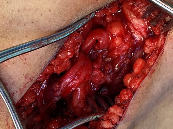
Figure 2. In the same patient as in Figure 1, the peroneal component of the sciatic nerve was coapted to the motor branch of the semimembranosus muscle with interrupted 6-0 prolene sutures.
The authors’ preferred technique for a BKA includes the use of a fishmouth incision with a posterior myocutaneous flap as described by the Attinger group[61]. In brief, the ideal tibial osteotomy is marked on the anterior leg approximately 4 finger breadths or 10-15 cm from the tibial tubercle. The fibular osteotomy should be 1-2 cm shorter than the tibial osteotomy. The anterior skin incision is made 1 cm distal to the planned tibial osteotomy. This line is carried through the lateral axis of the calf, which is defined as connecting the lateral femoral condyle to the lateral malleolus. Posteriorly, the myocutaneous flap is taken distally at the level of the Achilles tendon and trimmed back as needed prior to final closure[61]. The saphenous, sural, superficial peroneal, deep peroneal, and tibial nerves are identified. Common innervation muscle entry points (MEPs) have been defined by previous anatomical studies[55]. In the lateral compartment, the peroneus brevis and longus have MEPs on the medial and deep aspects. In the anterior compartment, the extensor digitorum longus has a readily identifiable MEP on the medial aspect of the muscle. In the posterior compartment, small branches of the tibial nerve can be found by following this nerve. While these branches can often be found by gross examination and knowledge of known anatomical MEPs, the authors utilize the Checkpoint Stimulator to confirm their neural characteristic and TMR is performed with several interrupted 6-0 to 8-0 prolene sutures, depending on the size of the nerves. This coaptation can be reinforced with Tisseel fibrin glue (Baxter International; Deerfield, IL). When nerve targets are not easily identifiable, then the authors quickly turn to RPNI using extraneous nearby muscles as grafts. This technique is described in detail below.
Outcomes of TMR [Table 1]
Despite initial concerns that the nerve transfer may cause new neuropathic pain, most patients find significant improvement in pain with minimal risk. Intraoperatively, identification of the motor entry points is the longest part of this procedure, but facilitated by previously published anatomic guides as well as the use of nerve stimulators. Coaptation of the nerves themselves is quite easily and quickly done, especially by those trained in microsurgery.
Outcomes research on TMR is still ongoing, but results have been favorable for pain management thus far. Dumanian et al.[27] conducted a prospective, single-blinded, randomized clinical trial at two centers and compared TMR to standard treatment with neuroma excision and burial of the nerve. Twenty-eight major upper and lower limb amputees with neuroma pain were randomized to these two groups. They conducted pain measures with two patient-reported scales, including the numerical rating scale (NRS) and PROMIS pain behavior, intensity, and interference short surveys. They also performed MRI neurograms and functional outcome assessment with the neuro-quality of life (neuro-QOL) measure. At one-year, residual limb and phantom limb pains trended toward improvement with TMR compared to standard treatment, although it did not reach statistical significance. Seventy-two percent of the TMR patients had no or mild phantom limb pain, and 67% had no or mild residual limb pain. Postoperative MRI nerve volumes were smaller for TMR patients. However, there was little difference in the neuro-QOL functional outcomes in this study[27]. Mioton et al.[62] performed a prospective study of 33 upper and lower extremity amputees from 2013-2017. Patient-reported outcomes measures with both NRS and PROMIS pain behavior, intensity, and interference scores demonstrated a significant decrease in residual limb and phantom limb pains at one-year follow-up compared to preoperatively. Quality of life and functional assessment measures were also significantly improved after TMR for both upper and lower extremity amputees. The authors recommend TMR as the first-line surgical treatment option for chronic amputation-related pain[62]. Bowen et al.[63] are performing an ongoing prospective multicenter, randomized study comparing the long-term success of TMR with the current standard of care. Preliminary results demonstrated that neuroma pain and PLP might persist immediately after surgery, but by 3-6 months post-operation, patients can expect reduced pain, less inhibition from PLP, and improvement in function and prosthesis use[63].
Effective treatment of existing symptomatic neuroma with TMR led teams across the country to assess its effect on neuroma pain prophylaxis by incorporating it in the index amputation surgery. Researchers have hypothesized that primary TMR has the advantages of decreased muscle atrophy due to less muscle denervation, improved nerve in-growth, and greater neuroplasticity[63]. Thus far, primary TMR has demonstrated promising results. The first report of TMR in the acute setting at the time of traumatic shoulder disarticulation demonstrated multiple successful nerve transfers for myoelectric prosthesis control as well as a complete lack of neuroma pain on the exam, which was corroborated with PROMIS at 8 months post-operation[64]. The patient did not use a prosthesis due to financial reasons, but distinct myoelectric control sites were seen on a clinical exam when she was able to trigger contraction under cortical control[64]. She did, however, report phantom sensations without pain, functional limitations, and mood depression[64]. In another study by Alexander et al.[65], patients who underwent major limb amputation for oncologic purposes with TMR at the time of amputation were compared to a cross-sectional sample of oncologic amputees who were not treated at the research institution (who therefore presumably had amputations without TMR). At one year follow-up, those who had primary TMR had significantly less frequent and less intense neuroma symptoms[65]. Another multi-institutional cohort study of major limb amputees (due to any reason) compared 51 patients who had primary TMR to 438 amputees who did not have TMR[66]. Evaluation by Valerio et al.[66] at a median follow-up of 330 days demonstrated that those with primary TMR had significantly less residual limb and phantom limb pain than the control group. These results were corroborated by a recent cohort study of primary TMR patients. In a single surgeon, single-institution study, vasculopathic patients undergoing BKA were treated with primary TMR or traction neurectomy with muscle implantation[67]. One hundred primary TMR patients underwent TMR of the superficial peroneal nerve and tibial nerve and traction neurectomy with muscle implantation of the remaining major nerves. They were compared to one hundred patients who underwent only traction neurectomy and muscle implantation of all nerves. Baseline patient characteristics were similar, and at nearly 10-month follow-up of the primary TMR group and 18.5-month follow-up for the traction neurectomy group, pain, as defined by residual limb pain and phantom limb pain as well as represented by opioid use, was significantly better in the primary TMR group. In addition, ambulatory rates were significantly higher in the primary TMR group[67]. These results have supported multidisciplinary collaboration between plastic surgeons and surgical groups such as orthopedists and vascular surgeons who perform primary amputations at the time of the index operation. Ideally, this would reduce the need for subsequent operations and reduce total operative time and costs. At the authors’ institution, this multidisciplinary discussion has resulted in routine consult of the plastic surgeon to perform reconstructive lower extremity amputations in collaboration with the vascular or orthopedic teams, if not as the primary surgeon.
REGENERATIVE PERIPHERAL NERVE INTERFACE
History of RPNI
RPNI, similar to TMR, was also originally conceived as a biological interface to harness residual peripheral nerve signal to control neuroprosthetic devices, but was found incidentally to help with symptomatic neuroma pain treatment[68]. The RPNI construct consists of a residual peripheral nerve implanted into a free, autologous muscle graft after excision of the terminal neuroma bulb. The regenerating axons on the proximal nerve ending form new neuromuscular junctions on the denervated muscle graft[68]. Kung et al.[69] demonstrated this with both histology and EMG studies on a rat model in which the extensor digitorum longus (EDL) muscle was removed and used as free muscle grafts. These muscle transfers were later found to be successfully revascularized as well as reinnervated[69]. In another study, some rats underwent sham surgeries of exposure of the soleus muscle while others underwent division of the peroneal nerve and RPNI with or without neurotization[70]. At three months, the neurotized RPNI approached sham subjects in muscle action potential amplitude and area as well as motor unit numbers[70]. Another rat EDL model study by Frost et al.[71] demonstrated that EMG signals could be acquired from RPNIs and translated into real-time, proportional control of neuroprosthetic hands with reduced signal contamination compared to control groups. Implantation of microscale electrodes either within the nerves themselves[72] or within the RPNI muscle grafts[73] were both biocompatible with minimal signal noise in rat hind limb models. In these ways, RPNI bridges the signaling gap between a living peripheral nerve and a mechanical device and facilitates motor function and sensory feedback in prosthetic limbs[47].
Surgical technique of RPNI
Symptomatic neuromas are diagnosed in the usual fashion with history and exams. Intraoperatively, the neuroma is identified and excised. Larger caliber nerves such as the sciatic nerve may require intraneural dissection to isolate individual fascicles. Autologous muscle grafts are harvested from the amputated limb or nearby donor muscles. Grafts are harvested from healthy muscle in the direction of the fibers. Ideally, these grafts are 5-6 mm thick, and they should be without any atrophy, scar, tendons, or signs of infection. The proximal end of the transected nerve is placed parallel to the muscle fibers and secured typically with 6-0 nonabsorbable monofilament sutures in an epimysial-to-epineural fashion. These constructs are located away from the surgical incision and deep to any weight-bearing surface [Figure 3][68].
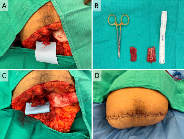
Figure 3. Reconstructive AKA with primary RPNI. A 75-year-old woman with a history of total left knee arthroplasty complicated by chronic peri-prosthetic infection who underwent multiple surgeries with poor bone and soft tissue availability underwent removal of hardware and left AKA. (A) The tibial and peroneal components of the sciatic nerve were dissected. (B) Small muscle grafts roughly
Creating an RPNI construct takes an estimated 7-10 min, according to the inventor group. Surgical creation of RPNI is technically simple and does not require expertise in microsurgery, unlike that with TMR. When, in TMR, the appropriate recipient motor nerves are difficult to identify, there is a limit on the number of independent muscle targets, or the patient is unable to tolerate a longer operation, RPNI is an excellent alternative option which can be elected for intraoperatively. This efficient, reproducible, and effective procedure has the potential to be implemented by surgeons from many disciplines with minimal additional cost[68] RPNI, like TMR, can be coded as pedicle nerve transfer (64905).
Outcomes of RPNI [Table 1]
Summary of clinical outcomes data in TMR and RPNI of the lower extremity
| Authors, year | Technique | Study design | Number of patients | Follow up | Outcomes |
| TMR | |||||
| Alexander et al.[65] 2019 | Primary TMR for upper and lower extremity oncologic amputation | Retrospective case series | 27: 6 upper extremity, 21 lower extremity | • Outcomes analyzed only for patients with greater than 1 year follow up • PROMIS pain intensity, behavior, and interference | • Significant improvement in mean PROMIS PLP • Significant improvement in mean PROMIS residual limb pain |
| Dumanian et al.[27] 2019 | Excision of post-amputation symptomatic neuroma with TMR | Prospective, single-blinded, randomized clinical trial | 28 (30 limbs total): 3 upper extremity TMR, 12 lower extremity TMR, 15 standard control | • Standard treatment patients in pain at 1 year were offered TMR • Pain measures with 2 patient-reported scales (NRS and PROMIS pain behavior, intensity, and interference) • MRI neurogram • Functional assessment with neuro-QOL | • Greater reduction in PLP and residual limb pain (not statistically significant) • 72% of TMR patients had no or mild PLP • 67% of TMR patients had no or mild residual limb pain • Little difference in neuro-QOL functional outcomes • Postoperative MRI nerve volumes are smaller for TMR than standard care patients |
| Valerio et al.[66] 2019 | Primary TMR or secondary TMR within 14 days of initial amputation | Retrospective multi-institutional cohort study | 51: 71% lower extremity 438 control major limb amputees | • Median follow up 330 days • Patient-reported outcomes (NRS and PROMIS pain behavior, intensity, and interference) | • PLP and residual limb pain scores lower with NRS and PROMIS in TMR group • Opioid prescription prevalence decreased postoperatively, especially for cancer patients |
| Mioton et al.[62] 2020 | Secondary TMR | Prospective cohort study | 33: 19 upper extremity, 14 lower extremity | • Outcomes analyzed after 1 year • Patient-reported outcomes (NRS and PROMIS pain behavior, intensity, and interference) • Functional assessment with OPUS for upper extremity, neuro-QOL for lower extremity | • PLP and residual limb pain decreased with NRS and PROMIS • OPUS scores increased • Neuro-QOL scores increased |
| Valerio et al.[76] 2020 | Primary (76%) and secondary (24%) TMR/vRPNI | Retrospective multi-institutional cohort study | 119 (123 limbs): 26 upper extremity, 93 lower extremity | • Follow up length not mentioned, but 8 lost, 111 patients actively participating currently | • One patient required secondary revision TMR/vRPNI for recurrent neuroma • 3 patients developed symptomatic neuromas in nerves that were not addressed |
| Chang et al.[67] 2021 | Primary TMR vs. traction neurectomy with muscle implantation in vasculopathic BKA | Retrospective single-surgeon, single-institution comparative cohort study | 100 primary TMR, 100 traction neurectomy with muscle implantation | • 9.6 months follow-up for primary TMR • 18.5 months follow-up for traction neurectomy • Binary pain assessment of residual limb, phantom limb, and opioid use • Binary assessment of ambulation | • Significant improvement in residual limb pain, phantom limb pain, and opioid use • Significant improvement in ambulation rates |
| RPNI | |||||
| Woo et al.[74] 2016 | Excision of post-amputation symptomatic neuroma with RPNI | Retrospective case series | 16: 3 upper extremity, 14 lower extremity | • Mean follow up 7.5 months • Postoperative phone interviews with patient-reported pains cores adapted from PROMIS • 100% response rate | • 81% able to wear prosthesis after RPNI (vs. 56% preoperatively) • Average pain score reduction 71% • 56% reduced pain medication use (none increased) • Aggregate pain interference score 2.15 (vs. 4.6 preoperatively) • 75% satisfied • 94% would have surgery again |
| Kubiak et al.[75] 2019 | Primary RPNI | Retrospective case series | 45 (52 limbs): 6 upper extremity, 46 lower extremity 45 control (52 limbs): 4 upper extremity, 48 lower extremity | • Mean follow up 357 days • Pain outcomes binary | • 13.3% of control patients developed symptomatic neuromas • 0% of primary RPNI patients developed symptomatic neuromas • 51% of primary RPNI patients developed PLP |
RPNI has demonstrated promising clinical results in improving neuroma pain and phantom pain.
As with TMR, primary RPNI at the time of amputation has also been increasingly explored with excellent preliminary results. A retrospective case series by Kubiak et al.[75] compared 45 patients who underwent primary RPNI, with both upper and lower extremity amputations to 45 control patients who underwent amputations without interfaces. Operative time was significantly longer in the primary RPNI group, with a mean of 152 min compared to 90 min in the control group who did not require identification of the major nerves. At a mean follow-up of nearly one year, those who had primary RPNI had a significantly fewer incidence of symptomatic neuroma and phantom limb pain. In fact, none of the primary RPNI patients developed symptomatic neuromas[75].
A combination technique of TMR with vascularized RPNI has also been trialed. In this technique,
CONCLUSION
Identification of the significant individual and systems burden of amputation, advances in microsurgery, as well as collaborative efforts with medicine-adjunct fields such as mechanical and bioengineering for prosthetics as well as neuromodulation are what birthed TMR and RPNI. These techniques to treat and, increasingly, prevent post-amputation pain are newer additions to the plastic surgeons’ armamentarium, which have allowed further evolution of the top rungs of the reconstructive ladder in an often challenging patient population[60].
Although these techniques were developed in the realm of plastic surgery, plastic surgery has a collaborative history in which many surgical interventions developed by plastic surgeons are adopted by other specialties. Amputation is performed by multiple surgical disciplines, including general surgery, orthopedic surgery, and vascular surgery, depending on the patient-specific indications as well as institution. Given the significant morbidity that post-amputation pain can have on these patients and the optimistic results with TMR and RPNI, collaboration with amputating surgeons is not only frontier medicine, but also owed to the patient to provide optimal care. In cases of limb salvage, the “orthoplastic approach” is the current gold standard[77]. Collaboration between orthopedists and plastic surgeons has been demonstrated to decrease time to definitive skeletal stabilization and soft tissue coverage, length of hospital stay, postoperative complications, and need for revision procedures, ultimately resulting in improved functional outcomes[78]. Amputation is an important alternative treatment for many in the same patient pool. One can conjecture that collaboration between plastic surgeons, and the primary amputating surgical specialties would result in improved outcomes, as demonstrated in preliminary results of primary TMR and RPNI.
Increasing evidence in the medical literature, as well as anecdotal evidence on a case-by-case basis, have made primary TMR and RPNI the gold standard at our institution and multi-surgical collaboration for these patients commonplace. Barriers to more widespread implementation of these new surgical techniques include awareness by the primary amputating surgeon as well as access to technical instruction[68]. Given how impactful these surgical interventions can be on amputees’ quality of life and function, inter-institutional conversation and live demonstration courses, among other strategies, are needed to increase access for this increasingly growing patient population.
DECLARATIONS
Authors’ contributionsMade substantial contributions to conception and design of the study, performed data analysis and interpretation, wrote and edited the manuscript: Toyoda Y, Azoury S, Bauder A, Levin LS, Kovach S
Availability of data and materialsNot applicable.
Financial support and sponsorshipNone.
Conflicts of interestStephen Kovach is a consultant for Checkpoint Surgical which is mentioned in the manuscript. Other authors declared that there are no conflicts of interest.
Ethical approval and consent to participateNot applicable.
Consent for publicationNot applicable.
Copyright© The Author(s) 2022.
REFERENCES
1. Hsu E, Cohen SP. Postamputation pain: epidemiology, mechanisms, and treatment. J Pain Res 2013;6:121-36.
2. Ephraim PL, Wegener ST, MacKenzie EJ, Dillingham TR, Pezzin LE. Phantom pain, residual limb pain, and back pain in amputees: results of a national survey. Arch Phys Med Rehabil 2005;86:1910-9.
3. Ziegler-Graham K, MacKenzie EJ, Ephraim PL, Travison TG, Brookmeyer R. Estimating the prevalence of limb loss in the United States: 2005 to 2050. Arch Phys Med Rehabil 2008;89:422-9.
4. Lo J, Chan L, Flynn S. A systematic review of the incidence, prevalence, costs, and activity and work limitations of amputation, osteoarthritis, rheumatoid arthritis, back pain, multiple sclerosis, spinal cord injury, stroke, and traumatic brain injury in the United States: a 2019 update. Arch Phys Med Rehabil 2021;102:115-31.
5. Chopra A, Azarbal AF, Jung E, et al. Ambulation and functional outcome after major lower extremity amputation. J Vasc Surg 2018;67:1521-9.
6. Bosse MJ, MacKenzie EJ, Kellam JF, et al. An analysis of outcomes of reconstruction or amputation after leg-threatening injuries. N Engl J Med 2002;347:1924-31.
7. Black CK, Ormiston LD, Fan KL, Kotha VS, Attinger C, Evans KK. Amputations versus salvage: reconciling the differences. J Reconstr Microsurg 2021;37:32-41.
8. Suckow BD, Goodney PP, Nolan BW, et al. Domains that determine quality of life in vascular amputees. Ann Vasc Surg 2015;29:722-30.
9. Sinha R, van den Heuvel WJ, Arokiasamy P. Factors affecting quality of life in lower limb amputees. Prosthet Orthot Int 2011;35:90-6.
10. Mundy LR, Klassen A, Grier AJ, et al. Identifying factors most important to lower extremity trauma patients: key concepts from the development of a patient-reported outcome instrument for lower extremity trauma, the LIMB-Q. Plast Reconstr Surg 2020;145:1292-301.
11. Döring K, Trost C, Hofer C, et al. How common are chronic residual limb pain, phantom pain, and back pain more than 20 years after lower limb amputation for malignant tumors? Clin Orthop Relat Res 2021;479:2036-44.
12. Amtmann D, Cook KF, Jensen MP, et al. Development of a PROMIS item bank to measure pain interference. Pain 2010;150:173-82.
13. Askew RL, Cook KF, Revicki DA, Cella D, Amtmann D. Evidence from diverse clinical populations supported clinical validity of PROMIS pain interference and pain behavior. J Clin Epidemiol 2016;73:103-11.
14. Srivastava D. Chronic post-amputation pain: peri-operative management - review. Br J Pain 2017;11:192-202.
15. Stankevicius A, Wallwork SB, Summers SJ, Hordacre B, Stanton TR. Prevalence and incidence of phantom limb pain, phantom limb sensations and telescoping in amputees: a systematic rapid review. Eur J Pain 2021;25:23-38.
16. Buch NS, Ahlburg P, Haroutounian S, Andersen NT, Finnerup NB, Nikolajsen L. The role of afferent input in postamputation pain: a randomized, double-blind, placebo-controlled crossover study. Pain 2019;160:1622-33.
17. Dellon AL. . Diagnosis and treatment of painful neuroma and of nerve compression in the lower extremity. 3rd ed. Elsevier Inc.; 2013.
19. Fuchs X, Flor H, Bekrater-Bodmann R. Psychological factors associated with phantom limb pain: a review of recent findings. Pain Res Manag 2018;2018:5080123.
20. Sood MK, Elliot D. Treatment of painful neuromas of the hand and wrist by relocation into the pronator quadratus muscle. J Hand Surg Br 1998;23:214-9.
21. Oliveira KMC, Pindur L, Han Z, Bhavsar MB, Barker JH, Leppik L. Time course of traumatic neuroma development. PLoS One 2018;13:e0200548.
22. Jong R, Shysh AJ. Development of a multimodal analgesia protocol for perioperative acute pain management for lower limb amputation. Pain Res Manag 2018;2018:5237040.
23. Finn SB, Perry BN, Clasing JE, et al. A randomized, controlled trial of mirror therapy for upper extremity phantom limb pain in male amputees. Front Neurol 2017;8:267.
24. Herrador Colmenero L, Perez Marmol JM, Martí-García C, et al. Effectiveness of mirror therapy, motor imagery, and virtual feedback on phantom limb pain following amputation: a systematic review. Prosthet Orthot Int 2018;42:288-98.
25. Pacheco-Barrios K, Meng X, Fregni F. Neuromodulation techniques in phantom limb pain: a systematic review and meta-analysis. Pain Med 2020;21:2310-22.
26. Eberlin KR, Ducic I. Surgical algorithm for neuroma management: a changing treatment paradigm. Plast Reconstr Surg Glob Open 2018;6:e1952.
27. Dumanian GA, Potter BK, Mioton LM, et al. Targeted muscle reinnervation treats neuroma and phantom pain in major limb amputees: a randomized clinical trial. Ann Surg 2019;270:238-46.
28. Oh C, Carlsen BT. New innovations in targeted muscle reinnervation: a critical analysis review. JBJS Rev 2019;7:e3.
29. Ives GC, Kung TA, Nghiem BT, et al. Current state of the surgical treatment of terminal neuromas. Neurosurgery 2018;83:354-64.
30. Stokvis A, van der Avoort DJC, van Neck JW, Hovius SER, Coert JH. Surgical management of neuroma pain: a prospective follow-up study. Pain 2010;151:862-9.
31. Guse DM, Moran SL. Outcomes of the surgical treatment of peripheral neuromas of the hand and forearm: a 25-year comparative outcome study. Ann Plast Surg 2013;71:654-8.
32. Domeshek LF, Krauss EM, Snyder-Warwick AK, et al. Surgical treatment of neuromas improves patient-reported pain, depression, and quality of life. Plast Reconstr Surg 2017;139:407-18.
33. Poppler LH, Parikh RP, Bichanich MJ, et al. Surgical interventions for the treatment of painful neuroma: a comparative meta-analysis. Pain 2018;159:214-23.
34. Souza JM, Purnell CA, Cheesborough JE, Kelikian AS, Dumanian GA. Treatment of foot and ankle neuroma pain with processed nerve allografts. Foot Ankle Int 2016;37:1098-105.
35. Economides JM, DeFazio MV, Attinger CE, Barbour JR. Prevention of painful neuroma and phantom limb pain after transfemoral amputations through concomitant nerve coaptation and collagen nerve wrapping. Neurosurgery 2016;79:508-13.
36. Prantl L, Schreml S, Heine N, Eisenmann-Klein M, Angele P. Surgical treatment of chronic phantom limb sensation and limb pain after lower limb amputation. Plast Reconstr Surg 2006;118:1562-72.
37. Sehirlioglu A, Ozturk C, Yazicioglu K, Tugcu I, Yilmaz B, Goktepe AS. Painful neuroma requiring surgical excision after lower limb amputation caused by landmine explosions. Int Orthop 2009;33:533-6.
38. Pet MA, Ko JH, Friedly JL, Smith DG. Traction neurectomy for treatment of painful residual limb neuroma in lower extremity amputees. J Orthop Trauma 2015;29:e321-5.
39. Kuiken TA, Li G, Lock BA, et al. Targeted muscle reinnervation for real-time myoelectric control of multifunction artificial arms. JAMA 2009;301:619-28.
40. Hijjawi JB, Kuiken TA, Lipschutz RD, Miller LA, Stubblefield KA, Dumanian GA. Improved myoelectric prosthesis control accomplished using multiple nerve transfers. Plast Reconstr Surg 2006;118:1573-8.
41. Kuiken TA, Barlow AK, Hargrove L, Dumanian GA. Targeted muscle reinnervation for the upper and lower extremity. Tech Orthop 2017;32:109-16.
42. Cheesborough JE, Smith LH, Kuiken TA, Dumanian GA. Targeted muscle reinnervation and advanced prosthetic arms. Semin Plast Surg 2015;29:62-72.
43. Kuiken TA, Miller LA, Lipschutz RD, et al. Targeted reinnervation for enhanced prosthetic arm function in a woman with a proximal amputation: a case study. Lancet 2007;369:371-80.
44. Dumanian GA, Ko JH, O’Shaughnessy KD, Kim PS, Wilson CJ, Kuiken TA. Targeted reinnervation for transhumeral amputees: current surgical technique and update on results. Plast Reconstr Surg 2009;124:863-9.
45. O’Shaughnessy KD, Dumanian GA, Lipschutz RD, Miller LA, Stubblefield K, Kuiken TA. Targeted reinnervation to improve prosthesis control in transhumeral amputees. A report of three cases. J Bone Joint Surg Am 2008;90:393-400.
46. Srinivasan SS, Diaz M, Carty M, Herr HM. Towards functional restoration for persons with limb amputation: a dual-stage implementation of regenerative agonist-antagonist myoneural interfaces. Sci Rep 2019;9:1981.
47. Kung TA, Bueno RA, Alkhalefah GK, Langhals NB, Urbanchek MG, Cederna PS. Innovations in prosthetic interfaces for the upper extremity. Plast Reconstr Surg 2013;132:1515-23.
48. Miller LA, Stubblefield KA, Lipschutz RD, Lock BA, Kuiken TA. Improved myoelectric prosthesis control using targeted reinnervation surgery: a case series. IEEE Trans Neural Syst Rehabil Eng 2008;16:46-50.
49. Zhou P, Lowery MM, Englehart KB, et al. Decoding a new neural machine interface for control of artificial limbs. J Neurophysiol 2007;98:2974-82.
50. Hargrove LJ, Miller LA, Turner K, Kuiken TA. Myoelectric pattern recognition outperforms direct control for transhumeral amputees with targeted muscle reinnervation: a randomized clinical trial. Sci Rep 2017;7:13840.
51. Agnew SP, Ko J, De La Garza M, Kuiken T, Dumanian G. Limb transplantation and targeted reinnervation: a practical comparison. J Reconstr Microsurg 2012;28:63-8.
52. Souza JM, Cheesborough JE, Ko JH, Cho MS, Kuiken TA, Dumanian GA. Targeted muscle reinnervation: a novel approach to postamputation neuroma pain. Clin Orthop Relat Res 2014;472:2984-90.
53. Kim PS, Ko JH, O'Shaughnessy KK, Kuiken TA, Pohlmeyer EA, Dumanian GA. The effects of targeted muscle reinnervation on neuromas in a rabbit rectus abdominis flap model. J Hand Surg Am 2012;37:1609-16.
54. Bowen JB, Ruter D, Wee C, West J, Valerio IL. Targeted muscle reinnervation technique in below-knee amputation. Plast Reconstr Surg 2019;143:309-12.
55. Fracol ME, Janes LE, Ko JH, Dumanian GA. Targeted muscle reinnervation in the lower leg: an anatomical study. Plast Reconstr Surg 2018;142:541e-50e.
56. Agnew SP, Schultz AE, Dumanian GA, Kuiken TA. Targeted reinnervation in the transfemoral amputee: a preliminary study of surgical technique. Plast Reconstr Surg 2012;129:187-94.
57. Gart MS, Souza JM, Dumanian GA. Targeted muscle reinnervation in the upper extremity amputee: a technical roadmap. J Hand Surg Am 2015;40:1877-88.
58. Serino A, Akselrod M, Salomon R, et al. Upper limb cortical maps in amputees with targeted muscle and sensory reinnervation. Brain 2017;140:2993-3011.
59. Morgan EN, Kyle Potter B, Souza JM, Tintle SM, Nanos GP 3rd. Targeted muscle reinnervation for transradial amputation: description of operative technique. Tech Hand Up Extrem Surg 2016;20:166-71.
60. Azoury SC, Bauder A, Souza JM, et al. Expanding the top rungs of the extremity reconstructive ladder: targeted muscle reinnervation, osseointegration, and vascularized composite allotransplantation. Plast Aesthet Res 2020;7:4.
61. Brown BJ, Iorio ML, Klement M, et al. Outcomes after 294 transtibial amputations with the posterior myocutaneous flap. Int J Low Extrem Wounds 2014;13:33-40.
62. Mioton LM, Dumanian GA, Shah N, et al. Targeted muscle reinnervation improves residual limb pain, phantom limb pain, and limb function: a prospective study of 33 major limb amputees. Clin Orthop Relat Res 2020;478:2161-7.
63. Bowen JB, Wee CE, Kalik J, Valerio IL. Targeted muscle reinnervation to improve pain, prosthetic tolerance, and bioprosthetic outcomes in the amputee. Adv Wound Care (New Rochelle) 2017;6:261-7.
64. Cheesborough JE, Souza JM, Dumanian GA, Bueno RA Jr. Targeted muscle reinnervation in the initial management of traumatic upper extremity amputation injury. Hand (N Y) 2014;9:253-7.
65. Alexander JH, Jordan SW, West JM, et al. Targeted muscle reinnervation in oncologic amputees: early experience of a novel institutional protocol. J Surg Oncol 2019;120:348-58.
66. Valerio IL, Dumanian GA, Jordan SW, et al. Preemptive treatment of phantom and residual limb pain with targeted muscle reinnervation at the time of major limb amputation. J Am Coll Surg 2019;228:217-26.
67. Chang BL, Mondshine J, Attinger CE, Kleiber GM. Targeted muscle reinnervation improves pain and ambulation outcomes in highly comorbid amputees. Plast Reconstr Surg 2021;148:376-86.
68. Kubiak CA, Kemp SWP, Cederna PS. Regenerative peripheral nerve interface for management of postamputation neuroma. JAMA Surg 2018;153:681-2.
69. Kung TA, Langhals NB, Martin DC, Johnson PJ, Cederna PS, Urbanchek MG. Regenerative peripheral nerve interface viability and signal transduction with an implanted electrode. Plast Reconstr Surg 2014;133:1380-94.
70. Urbanchek MG, Baghmanli Z, Moon JD, Sugg KB, Langhals NB, Cederna PS. Quantification of regenerative peripheral nerve interface signal transmission. Plast Reconstr Surg 2012;130:55-6.
71. Frost CM, Ursu DC, Flattery SM, et al. Regenerative peripheral nerve interfaces for real-time, proportional control of a Neuroprosthetic hand. J Neuroeng Rehabil 2018;15:108.
72. Urbanchek MG, Wei B, Egeland BM, Abidian MR, Kipke DR, Cederna PS. Microscale electrode implantation during nerve repair: effects on nerve morphology, electromyography, and recovery of muscle contractile function. Plast Reconstr Surg 2011;128:270e-8e.
73. Ursu DC, Urbanchek MG, Nedic A, Cederna PS, Gillespie RB. In vivo characterization of regenerative peripheral nerve interface function. J Neural Eng 2016;13:026012.
74. Woo SL, Kung TA, Brown DL, Leonard JA, Kelly BM, Cederna PS. Regenerative peripheral nerve interfaces for the treatment of postamputation neuroma pain: a pilot study. Plast Reconstr Surg Glob Open 2016;4:e1038.
75. Kubiak CA, Kemp SWP, Cederna PS, Kung TA. Prophylactic regenerative peripheral nerve interfaces to prevent postamputation pain. Plast Reconstr Surg 2019;144:421e-30e.
76. Valerio I, Schulz SA, West J, Westenberg RF, Eberlin KR. Targeted muscle reinnervation combined with a vascularized pedicled regenerative peripheral nerve interface. Plast Reconstr Surg Glob Open 2020;8:e2689.
77. Levin LS. The reconstructive ladder. An orthoplastic approach. Orthop Clin North Am 1993;24:393-409.
Cite This Article
Export citation file: BibTeX | RIS
OAE Style
Toyoda Y, Azoury S, Bauder A, Levin LS, Kovach S. Lower extremity amputation: the emerging role of targeted muscle reinnervation (TMR) and regenerative peripheral nerve interface (RPNI). Plast Aesthet Res 2022;9:17. http://dx.doi.org/10.20517/2347-9264.2021.118
AMA Style
Toyoda Y, Azoury S, Bauder A, Levin LS, Kovach S. Lower extremity amputation: the emerging role of targeted muscle reinnervation (TMR) and regenerative peripheral nerve interface (RPNI). Plastic and Aesthetic Research. 2022; 9: 17. http://dx.doi.org/10.20517/2347-9264.2021.118
Chicago/Turabian Style
Toyoda, Yoshiko, Said Azoury, Andrew Bauder, L. Scott Levin, Stephen Kovach. 2022. "Lower extremity amputation: the emerging role of targeted muscle reinnervation (TMR) and regenerative peripheral nerve interface (RPNI)" Plastic and Aesthetic Research. 9: 17. http://dx.doi.org/10.20517/2347-9264.2021.118
ACS Style
Toyoda, Y.; Azoury S.; Bauder A.; Levin LS.; Kovach S. Lower extremity amputation: the emerging role of targeted muscle reinnervation (TMR) and regenerative peripheral nerve interface (RPNI). Plast. Aesthet. Res. 2022, 9, 17. http://dx.doi.org/10.20517/2347-9264.2021.118
About This Article
Special Issue
Copyright

Data & Comments
Data


 Cite This Article 23 clicks

 Like This Article 24
likes
Like This Article 24
likes


Comments
Comments must be written in English. Spam, offensive content, impersonation, and private information will not be permitted. If any comment is reported and identified as inappropriate content by OAE staff, the comment will be removed without notice. If you have any queries or need any help, please contact us at support@oaepublish.com.