
Free tissue reconstruction of massive facial trauma - review of the literature and considerations to implement aesthetic and functional outcome
 0
0Abstract
The surgical armamentarium for the treatment of massive facial trauma has undergone a dramatic shift from early management strategies. Although tenants of acute trauma management continue to prioritize airway management and cardiopulmonary support, improved functional outcomes are achievable with an emphasis on early definitive free tissue transfer. The use of workhorse donor flaps, such as the radial forearm, fibula, and latissimus, have become the standard of care. An emphasis is placed on the separation of cranial, sinonasal, and oral contents and restoration of form and function. Here, we also discuss the management of telecanthus, nasal defects, and microstomia - sequelae which represent unique challenges to the reconstructive surgeon. The ability to perform virtual surgical planning and facial transplantation will likely shape future paradigms and represent the need to perform ongoing research.
Keywords
INTRODUCTION
Massive facial trauma presents a historically complex problem for patients and those charged with management and reconstruction in this setting. The patient must first be stabilized at initial presentation, after which attention may then be turned towards questions regarding the nature of the injury itself and the ideal plan for restoration of form and function. Facial trauma represents a substantial burden on the medical system with estimated costs of around $1 billion annually, with a subset of this cost related to the care of patients with massive facial trauma[1]. Although not technically defined, massive facial trauma can be understood as a facial injury, more often due to ballistic or avulsive forces, that result in significant soft-tissue and bone loss necessitating substitution of missing components through a variety of surgical practices[2]. Technological advances in the last century, however, have not only given surgeons new techniques for improved reconstruction but have also contributed to changes in the pattern of injury over time.
TRENDS IN PATTERNS OF INJURY
There is a paucity of large-scale studies regarding the epidemiology and treatment of massive facial trauma patients; however, inferences can be made by combining related trends in the literature with anecdotal and institutional experience. Mechanisms typically capable of producing such injuries are gunshot injuries or, less likely, high-speed motor-vehicle accidents. Although motor-vehicle usage has become widespread throughout the 20th century, advanced safety measures such as seatbelts, airbags, and vehicle design have led to decreased incidence of associated facial injuries in several studies[3,4]. Ballistic injuries to the face are much more likely to produce injuries classifiable as massive facial trauma, and the United States continues to suffer from an epidemic of gun violence, as evidenced by the roughly eight times higher death rate due to firearms than other high-income nations[5,6]. More recent evidence alarmingly shows an increase in age-adjusted rates of firearm mortality in the United States from 2014 to 2017 after having been stable for nearly a decade prior[7]. Among the roughly 329 daily injuries related to firearms, two-thirds of patients will survive and go on to live with the functional ramifications of their injuries[8].
TRENDS IN RECONSTRUCTIVE PRACTICE
Mechanisms capable of generating massive facial trauma wounds impart a number of locoregional tissue changes that create challenges for definitive primary repair, including evolving tissue loss, distortion of normal landmarks, muscular attachment disruption, and an overall deficit of tissue bulk, both soft tissue and bone[2,9-11]. Prior to the implementation of free tissue transfer, non-vascularized bone grafts were the primary technique used in order to restore the rigid facial structure on top of which soft tissues could be manipulated. These efforts were met with significant issues, among which were resorption and relative the inability to reconstruct larger defects due to their lack of vascularity[12]. Throughout the period surrounding World War I and II, attempts were made to incorporate bone grafts into random pattern neck skin flaps as a method of enhancing reconstructive efforts[13]. This would eventually give way to the use of pedicled myocutaneous flaps in the head and neck; however, limitations due to the reach of the flap and the sub-optimal transfer of bone were still problematic[14]. Ultimately, the fibula free flap would be described as the first free vascularized bone flap in 1975 and was readily incorporated into the reconstruction of the head and neck after that[15]. Numerous other osseus flaps have since been described and added to the armamentarium of the modern microvascular surgeon. This has led to a shift in the paradigm of management for massive facial trauma patients from delayed to more definitive primary reconstructive efforts. As will be described in this paper, modern approaches aim to overcome the pitfalls of delayed management by decreasing wound contracture and scarring before they can further distort the complexities of the facial anatomy.
PHASED APPROACH
Phase I
Futran et al.[2] (senior author) proposed a three-phased approach emphasizing early definitive reconstruction when possible (see Figure 1). Over time, early reconstruction has gained favor over delayed reconstruction. Many advantages include minimization of scar contractures of facial soft tissues, reduced infection risk, and improved long-term function[2,16-19]. Such scar contractures and distortion of underlying bony architecture render an acceptable cosmetic and functional result nearly impossible. Previously, the senior author reported on a case series of 49 patients treated with an approach emphasizing early reconstruction undergoing free tissue transfer for severe facial trauma with excellent success rates - no flap failures, excellent/very good cosmesis, and 98% per oral alimentation without the need for gastrostomy tube feeding[2].
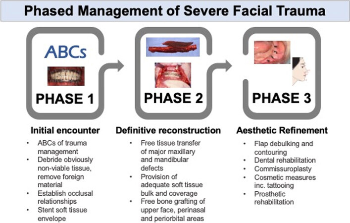
Figure 1. Three phases of management of severe facial trauma. Phase 1 emphasizes stabilization and preparation for definitive reconstruction with free tissue transfer for major defects in Phase 2, followed by adjunctive procedures for aesthetic refinement in Phase 3.
The goal of phase I is to manage life-threatening injuries in accordance with the ABCs of emergency care and supported by Advanced Trauma Life Support protocols. Securing the airway, controlling bleeding, and managing shock is of paramount importance as the focus is on the stabilization of the patient for imaging and initial operative management. From an operative standpoint, the goals of phase I are to:
(1) Assess intracranial exposure, orbital injury, or carotid exposure for immediate coverage[16];
(2) Establish occlusal relationships with maxillomandibuar fixation with or without dental splinting;
(3) Debride foreign material obviously non-viable tissue; questionably viable tissue should be left for reassessment in 24-72 h;
(4) Stenting of the soft tissue envelope to prevent contracture (free bone grafts vs. reconstruction plates across segmental mandibular defects);
(5) Assess anatomic deficits for the planning of definitive repair;
(6) Optionally, stent nasolacrimal and parotid ducts for eventual definitive repair.
Additionally, patient education and expectation setting are critical.
Phase II
In phase II, early definitive reconstruction is performed. Major upper-face, mid-face, and mandibular defects are reconstructed with appropriately selected donor tissues. Non-vascularized cranial bone grafts are especially useful in the upper face but are contra-indicated if overlying soft tissue is inadequate. Commonly used donor sites for bony reconstruction of the mid- and lower-face include fibula, iliac crest, scapula, and radial forearm osteocutaneous free flaps. Some authors advocate for fibula and iliac crest over the scapula and radial forearm due to the possibility of placing osseointegrated dental implants, given that the trauma population tends to be young with a long life expectancy[19]. In addition, the long vascular pedicle of the fibula free flap is especially advantageous for mid-face reconstruction when a significant length is required to reach the recipient artery and veins of the neck.
Upper- and mid-face considerations
High-energy injuries to the upper- and mid-face can result in complex craniofacial, skull base, and orbital defects. Therefore, the need to separate exposed dura from the sinonasal cavity is of paramount importance[16]. Small to medium-sized cranial defects are amenable to pericranial-galeal flaps or rotational temporalis flaps. Larger skull base defects are well suited to reconstruction with the thin and broad latissimus muscle free flap covering custom implants from biocompatible materials such as titanium or polyetheretherketone[16].
Injuries to the periorbital and perinasal region disrupt sensitive anatomic structures, and resultant defects of the mid-face and orbital regions are challenging to reconstruct with local tissues, as rotational flap options are limited. This area is also critical to the foundation and perception of personal identity, as the collective aesthetic relationships and contours of the eyes, nose, and lips are central to facial recognition[10]. Free bone grafts can be utilized to provide the underlying framework of the orbital rims, recreate intercanthal distance, and provide an adequate projection of the reconstructed nose[9]. Intercanthal distance should be optimized in early definitive reconstruction, as delayed revision is particularly challenging. With the advent of virtual surgical planning and the ability to design patient-specific implants, biomaterials in this area will become a more prominent part of the surgical armamentarium.
For mid-face defects requiring soft tissue reconstruction alone, radial forearm or anterolateral thigh flaps provide adequate bulk and can be easily contoured at a later date. However, in massive facial trauma, there frequently exists the need to replace underlying bony architecture to restore facial projection, recreate facial buttresses, and resurface cutaneous as well as mucosal lining. Rodriguez et al.[19] presents a useful classification system for mid-face defects ranging from Class I (unilateral dentoalveolar defect) to Class IV (bilateral dentoalveolar defect plus orbital rim defects). The authors exclusively used fibula or iliac crest for maxillary reconstruction with the intention of future osseointegrated dental implants. In particular, for bilateral defects, the longer pedicle of the fibular free flap is advantageous. The iliac crest pedicle, which is shorter at 4-5 cm, is better suited to unilateral defects. A flexor hallucis longus or soleus muscle (fibula) or transversus abdominus muscle cuffs (iliac crest) may be used to resurface the intraoral lining.
Krane et al.[20] reported on three cases of using a single fibula osteocutaneous free flap to perform simultaneous maxillary and mandibular reconstruction. The neo-maxilla is fashioned from the distal bony segment and associated skin paddle. A segment of intervening bone is removed from the flap pedicle, and proximal bone is used to reconstruct the neo-mandible. Buccal adhesion or contracture is easily addressed with lysis of the offending adhesion.
Mandible considerations
Goals of mandibular reconstruction include the recreation of pre-morbid occlusion, closure and prevention of orocutaneous fistula, maintaining projection of the lower third of the mid-face, and preservation of intact nerves for optimal speech and swallow function. Occasionally, facial trauma may result in fractured but preserved mandibular bone attached to the overlying periosteum, which is amenable to rigid fixation without the need for additional free tissue. For example, such defects may occur in the setting of self-inflicted submental gunshot wounds[2].
The fibular osteocutaneous free flap has become the workhorse of mandibular reconstruction for segmental defects in the setting of trauma[2,21,22]. While non-vascularized bone grafts may be used for bony defects <
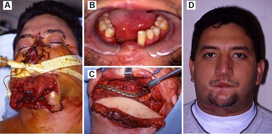
Figure 2. (A) A 31-year-old male presented with a gunshot wound to the oromandibular complex resulting in massive lower and midface trauma; (B) oral cutaneous fistula present in the anterior floor of mouth; (C) fibula free flap contoured to the defect for restoration of mandibular continuity; (D) 1-year post-injury result.
Phase III
In phase III, aesthetic contouring and functional refinement are performed. Soft tissue debulking, oral commissuroplasty, dental rehabilitation, tissue fillers, and facial contouring are undertaken. The timeline for adjunctive procedures is typically not within 6 months of the first definitive free tissue repair. Figure 3 illustrates successive procedures in a 19-year-old male who sustained a self-inflicted shotgun wound
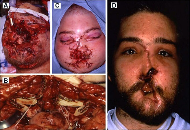
Figure 3. (A) A 23-year-old male who sustained a self-inflicted gunshot wound to the face, initial encounter; (B) ten days after the initial injury, repair of nasal Le Fort II fractures was performed with split calvarial bone grafts; (C) telecanthus as the result of migration of canthal tendons; (D) post-operative result after resuspension of medial canthi to screws placed into the frontal bone.
Telecanthus
Telecanthus results from severe upper- and mid-face trauma causing injury to the naso-orbito-ethmoid complex and disruption of the medial canthal tendon attachments. Kalavrezos et al.[25] defines telecanthus as having a canthal index (ratio of intercanthal distance to outercanthal distance × 100) of 38. The fine contours of the medial palpebral fissure nasal prominence, and naso-orbital angels are subsequently lost[26]. Gold standard treatment is transnasal wiring of disrupted medial canthal tendons, but even then, the incidence of post-operative telecanthus can be considerable, up to 14% in some series[25]. This is due to the lateral forces exerted on by contracting tissues of the NOE region, and authors often advocate for miniplate fixation of the comminuted NOE complex with an emphasis on overcorrection[27].
Figure 3B demonstrates principles of NOE trauma management as well as challenges of telecanthus prevention. Mini-plate fixation of Lefort II and III fractures using split calvarial bone grafts with the wiring of the bilateral avulsed medial canthal tendons was performed ten days after the initial injury. The pre-operative intercanthal distance was 58 mm. Subsequently, transnasal wiring was performed, pulling the medial canthal tendons superoposteriorly with an overcorrection. Over the ensuing month, there was the migration of the canthal tendons resulting in telecanthus [Figure 3C], and they were resuspended using screws placed into the frontal bone, resulting in improvement of intercanthal distance Figure 3D. However, over the ensuing year and a half later, the wires pulled through the soft tissues, and the patient had relaxion of his repair, and twice he required narrowing of the nasal region and resuspension of bilateral canthi to frontal screws.
Microstomia
Oral incompetence and microstomia may manifest after severe facial trauma, often as the result of late sequelae and scarring. The reader is directed towards more comprehensive sources reviewing contemporary perioral reconstruction[28]; however, several key principles deserve attention. Firstly, restoring dynamic motion and sensation is paramount. To that end, local reconstructive techniques, including cheek advancement techniques, Karapandzic flaps [Figure 4A-D], and commisuroplasties, are highly effective in restoring defects of less than 80% of the lips[29-32]. Hanasono and Langstein[30] report the extension of the traditional Karapandzic flap technique to include tissue from the perioral cheek, allowing near-total lower lip defects to be reconstructed with sensation and dynamic function. When local flaps are insufficient, the radial forearm free flap has traditionally been the most favorable donor site given its thinness and pliability. Modifications, including the palmaris longus tendon to create a more durable sling, have also been reported[33]. More recently, the gracilis muscle free flap has been used to create a neo-sphincter, with reinnveration by anastomosis to the marginal mandibular nerve. Gurunluoglu et al.[34] demonstrate that the gracilis free flap is useful for lower lip defects in ballistic injuries in particular, with 3/3 patients regaining speech and oral competence. Subsequent adjunctive procedures, including vermillion tattoo and mucosal advancements, may be necessary.
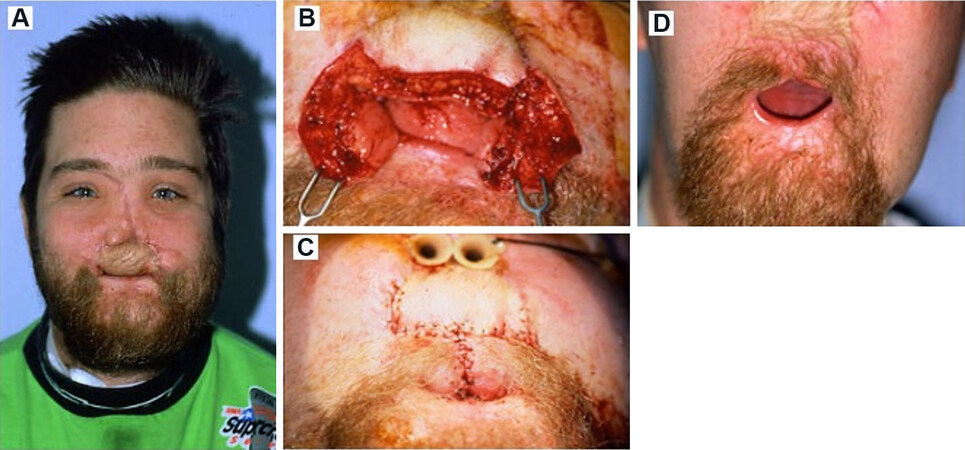
Figure 4. (A) The patient in Figure 3 underwent a radial forearm osteocutaneous free flap for maxillary reconstruction 6 months post-injury but continued to have microstomia. (B, C) He subsequently underwent upper lip reconstruction with bilateral Karapandzic flaps. (D) Post-operative result after bilateral Karapandzic flaps.
Total nasal reconstruction
The nose is often considered the focal point of the face with both important aesthetic and functional concerns, which requires careful forethought when planning for total nasal reconstruction. Appropriate management aims to recapitulate nasal projection and ideally restore nasal airflow while avoiding paranasal sinus obstruction, but the complex three-dimensional shape of the native nose, as well as the multiple tissue types involved, create hurdles for achieving ideal outcomes. An algorithm for nasal reconstruction was proposed by Manson et al.[35] in 1979 and remains useful to this day. A variety of techniques are able to be employed depending on the nature of the defect and whether cutaneous, support or nasal lining structure is missing from the defect. In cases of total nasal reconstruction, there has been successfully combining the paramedian forehead flap for cutaneous reconstruction with the radial forearm flap for nasal lining[36]. Recreating support structures for these defects maintains projection and contour within the reconstructed soft tissue envelope but requires cantilevered bone grafting in situations where the nasal bones are lost due to injury[37].
The patient from Figures 3 and 4 suffered from full-thickness deformity of all nasal subunits. After underlying framework repair with a fibular free flap and radial forearm osteocutaneous free flap for the mandible and maxilla, respectively, the patient continued to have a noticeable premaxillary deficiency, lacked any nasal projection, and had a nonexistent nasal airway [Figure 5A]. The patient then underwent total nasal reconstruction. First, internal lining replacement was achieved through turn-in mucosal advancement and septal mucosal flaps [Figure 5B]. Then, cranial bone grafts were harvested from the temporal bones and fixed to the nasofrontal angle [Figure 5C and D].
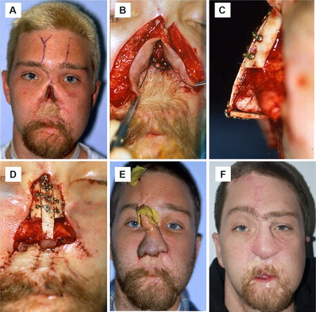
Figure 5. (A) Two years after the initial injury, the aforementioned patient persisted in having a total nasal defect with the absence of all nasal subunits; (B) turn-in mucosal advancement flaps were performed to recreate the nasal lining; (C, D) total nasal reconstruction was performed with calvarial free bone grafts to create underlying nasal framework; (E) a paramedian forehead flap was performed for soft tissue reconstruction of the external nose; (F) final reconstructive result at 3.5 years post-injury.
Cutaneous reconstruction required initial paramedian forehead flap usage [Figure 5E] with a subsequent revision paramedian forehead flap employed after a frontal tissue expander was placed. Support of the nose was achieved with initial attempts at autogenous cartilage grafting but ultimately required placement of a cantilevered split calvarial bone graft underneath the second paramedian forehead flap to maintain projection. Employing these techniques together over a period of approximately 4 years, the final nasal reconstruction demonstrates improved projection, maintenance of bilateral nasal airways, and adequate symmetry [Figure 5F].
NEW ADVANCES
Virtual surgical planning
Virtual surgical planning (i.e., VSP) has grown in popularity for the reconstruction of the head and neck region. Elimination of intra-operative guesswork compared to free-handed approaches and increased reconstructive precision are possible. While no technology can supplant experience, VSP has the potential to assist more novice surgeons in tackling the reconstructive challenge of complex craniofacial defects. Stranix et al.[38] presented two solutions to not having a pre-operative CT scan for planning purposes: using a mirror-image reconstruction of the uninvolved side or using normative data in lieu of an intact contralateral side.
Initial results with VSP-aided free flap reconstruction of the traumatized face have been encouraging.
Facial transplantation
One major limitation of autologous free tissue transfer is the satisfactory reconstruction of the central face and mimetic structures such as the lips and eyelids. Nasal defects can be satisfactorily reconstructed with a paramedian forehead flap, but local and rotational flaps lack the delicate pliability needed to reconstruct the periorbita, including eyelids. Moreover, the central face and the relative relationships and contours of the eyes, nose, and lips are described by Alam and Chi[10] as being the most key to personal identity. Relative indications for facial transplantation have been stated as being defects that are severe and involved the central face, including both upper and lower lids and upper and lower lips[42].
To that end, facial transplantation has evolved as a last-resort option to reconstruct defects from massive facial trauma failing free flap repair. Indeed, the first face transplant in the United States was performed after the patient had undergone 23 prior procedures and 4 failed free flaps. Since then, more than 40 facial transplants have been performed worldwide. The vast majority of such injuries have been due to ballistic, thermal burn, blunt force, or animal-related facial traumas[43]. Short-term outcomes have been excellent, with full graft take, recovery of sensory and motor nerve function, and improved cosmesis and quality of life being the norm[10,42,43]. Although 10-year allograft survival exceeds 80%[44], the greatest challenge is the need for lifelong triple-drug immunosuppression. Acute rejection often responds to steroid therapy or plasmapheresis, but chronic rejection has been more difficult to treat, and chronic antibody-mediated rejection has resulted in graft loss in several patients. One of these was a patient with neurofibromatosis who suffered complete graft loss at year 7 but became the first successful facial retransplantation[45]. He was treated with an aggressive immune desensitization regimen and endured a 1-year post-retransplantation hospitalization but has enjoyed graft survival > 30 months after retransplantation.
CONCLUSION
Severe facial trauma continues to be a significant health burden. After stabilization of the initial injury, early definitive free tissue transfer performed after 72 h results in improved function and cosmesis. Special considerations must be taken to the closure of intracranial defects and the separation of sinonasal and oropharyngeal cavities. Additional secondary aesthetic restoration procedures are often necessary. For severe central face defects, facial transplantation is evolving as a viable option.
DECLARATIONS
Authors’ contributionsMade substantial contributions to conception and literature review: Pang J, Cash H
Oversaw manuscript structure and content with provision of case details: Futran N
Availability of data and materialsNot applicable.
Financial support and sponsorshipNone.
Conflicts of interestThe senior author Futran N is an educational consultant for Stryker Corp.
Ethical approval and consent to participateNot applicable.
Consent for publicationThe study is obtained the consent from patients.
Copyright© The Author(s) 2021.
REFERENCES
1. Allareddy V, Allareddy V, Nalliah RP. Epidemiology of facial fracture injuries. J Oral Maxillofac Surg 2011;69:2613-8.
2. Futran ND, Farwell DG, Smith RB, Johnson PE, Funk GF. Definitive management of severe facial trauma utilizing free tissue transfer. Otolaryngol Head Neck Surg 2005;132:75-85.
3. Roden KS, Tong W, Surrusco M, Shockley WW, Van Aalst JA, Hultman CS. Changing characteristics of facial fractures treated at a regional, level 1 trauma center, from 2005 to 2010: an assessment of patient demographics, referral patterns, etiology of injury, anatomic location, and clinical outcomes. Ann Plast Surg 2012;68:461-6.
4. VandeGriend ZP, Hashemi A, Shkoukani M. Changing trends in adult facial trauma epidemiology. J Craniofac Surg 2015;26:108-12.
5. Tasigiorgos S, Economopoulos KP, Winfield RD, Sakran JV. Firearm injury in the United States: an overview of an evolving public health problem. J Am Coll Surg 2015;221:1005-14.
6. Ranney ML, Sankoff J, Newman DH, et al. A call to action: firearms, public health, and emergency medicine. Ann Emerg Med 2013;61:700-2.
7. Goldstick JE, Zeoli A, Mair C, Cunningham RM. US firearm-related mortality: national, state, and population trends, 1999-2017. Health Aff (Millwood) 2019;38:1646-52.
8. Kaufman EJ, Wiebe DJ, Xiong RA, Morrison CN, Seamon MJ, Delgado MK. Epidemiologic trends in fatal and nonfatal firearm injuries in the US, 2009-2017. JAMA Intern Med 2021;181:237-44.
9. Kim RY, Sokoya M, Williams FC, Shokri T, Ducic Y. Role of free tissue transfer in facial trauma. Facial Plast Surg 2019;35:584-9.
10. Alam DS, Chi JJ. Facial transplantation for massive traumatic injuries. Otolaryngol Clin North Am 2013;46:883-901.
11. Clark N, Birely B, Manson PN, et al. High-energy ballistic and avulsive facial injuries: classification, patterns, and an algorithm for primary reconstruction. Plast Reconstr Surg 1996;98:583-601.
12. Sparks DS, Saleh DB, Rozen WM, Hutmacher DW, Schuetz MA, Wagels M. Vascularised bone transfer: history, blood supply and contemporary problems. J Plast Reconstr Aesthet Surg 2017;70:1-11.
13. Blair VP. Surgery and diseases of the mouth and jaws. 3th ed. London: Henry Kimpton; 1918. p. 153-4.
14. Biller HF, Baek SM, Lawson W, Krespi YP, Blaugrund SM. Pectoralis major myocutaneous island flap in head and neck surgery: analysis of complications in 42 cases. Arch Otolaryngol 1981;107:23-6.
15. Taylor GI, Miller GD, Ham FJ. The free vascularized bone graft. A clinical extension of microvascular techniques. Plast Reconstr Surg 1975;55:533-44.
16. Funk GF, Laurenzo JF, Valentino J, McCulloch TM, Frodel JL, Hoffman HT. Free-tissue transfer reconstruction of midfacial and cranio-orbito-facial defects. Arch Otolaryngol Head Neck Surg 1995;121:293-303.
17. Zeiderman MR, Pu LLQ. Contemporary reconstruction after complex facial trauma. Burns Trauma 2020;8:tkaa003.
18. McDonald TJ, Lopez MA. Management of facial trauma: lessons of war. Facial Plast Surg 2010;26:482-7.
19. Rodriguez ED, Martin M, Bluebond-Langner R, Khalifeh M, Singh N, Manson PN. Microsurgical reconstruction of posttraumatic high-energy maxillary defects: establishing the effectiveness of early reconstruction. Plast Reconstr Surg 2007;120:103S-17S.
20. Krane NA, Fagin A, Ghanem TA, Cannady SB, Petrisor D, Wax MK. Simultaneous maxillary and mandibular reconstruction with a single Osteocutaneous fibula free flap: a description of three cases. Microsurgery 2021;41:79-83.
21. Gurunluoglu R, Gatherwright J. Microsurgical reconstruction of complex maxillofacial gunshot wounds: outcomes analysis and algorithm. Microsurgery 2019;39:384-94.
22. Eser C, Gencel E, Kesiktaş E, Yavuz M. Outcomes of anatomic reconstruction of gunshot-inflicted lower face defects by free Osteoseptocutaneous fibula flap and expanded or nonexpanded temporal scalp flap combination in males. J Craniofac Surg 2016;27:1139-42.
23. Chattha A, Lee JC, Johnson PK, Patel A. An Algorithmic Approach to the management of ballistic facial trauma in the civilian population. J Craniofac Surg 2018;29:2010-6.
24. Wei FC, Seah CS, Tsai YC, Liu SJ, Tsai MS. Fibula osteoseptocutaneous flap for reconstruction of composite mandibular defects. Plast Reconstr Surg 1994;93:294-304; discussion 5-6.
25. Kalavrezos ND, Graetz KW, Eyrich GK, Sailer HF. Late sequelae after high midface trauma. J R Coll Surg Edinb 2000;45:359-62.
26. Elbarbary AS, Ali A. Medial canthopexy of old unrepaired naso-orbito-ethmoidal (noe) traumatic telecanthus. J Craniomaxillofac Surg 2014;42:106-12.
29. Mehra P, Caiazzo A, Bestgen S. Bilateral oral commissurotomy using buccal mucosa flaps for management of microstomia: report of a case. J Oral Maxillofac Surg 1998;56:1200-3.
30. Hanasono MM, Langstein HN. Extended Karapandzic flaps for near-total and total lower lip defects. Plast Reconstr Surg 2011;127:1199-205.
31. Berlet AC, Ablaza VJ, Servidio P. A refined technique for oral commissurotomy. J Oral Maxillofac Surg 1993;51:1400-3.
32. Ducic Y, Athre R, Cochran CS. The split orbicularis myomucosal flap for lower lip reconstruction. Arch Facial Plast Surg 2005;7:347-52.
33. Sadove RC, Luce EA, McGrath PC. Reconstruction of the lower lip and chin with the composite radial forearm-palmaris longus free flap. Plast Reconstr Surg 1991;88:209-14.
34. Gurunluoglu R, Glasgow M, Williams SA, Gurunluoglu A, Antrobus J, Eusterman V. Functional reconstruction of total lower lip defects using innervated gracilis flap in the setting of high-energy ballistic injury to the lower face: preliminary report. J Plast Reconstr Aesthet Surg 2012;65:1335-42.
35. Manson PN, Hoopes JE, Chambers RG, Jaques DA. Algorithm for nasal reconstruction. Am J Surg 1979;138:528-32.
36. Menick FJ, Salibian A. Microvascular repair of heminasal, subtotal, and total nasal defects with a folded radial forearm flap and a full-thickness forehead flap. Plast Reconstr Surg 2011;127:637-51.
37. Rohrich RJ, Griffin JR, Ansari M, Beran SJ, Potter JK. Nasal reconstruction--beyond aesthetic subunits: a 15-year review of 1334 cases. Plast Reconstr Surg 2004;114:1405-16; discussion 1417-9.
38. Stranix JT, Stern CS, Rensberger M, et al. A virtual surgical planning algorithm for delayed maxillomandibular reconstruction. Plast Reconstr Surg 2019;143:1197-206.
39. May MM, Howe BM, O'Byrne TJ, et al. Short and long-term outcomes of three-dimensional printed surgical guides and virtual surgical planning versus conventional methods for fibula free flap reconstruction of the mandible: decreased nonunion and complication rates. Head Neck 2021;43:2342-52.
40. Seruya M, Fisher M, Rodriguez ED. Computer-assisted versus conventional free fibula flap technique for craniofacial reconstruction: an outcomes comparison. Plast Reconstr Surg 2013;132:1219-28.
41. Navarro Cuéllar C, Martínez EB, Navarro Cuéllar I, et al. Primary maxillary reconstruction with fibula flap and dental implants: a comparative study between virtual surgical planning and standard surgery in Class IIC defects. J Oral Maxillofac Surg 2021;79:237-48.
43. Farber SJ, Kantar RS, Diaz-Siso JR, Rodriguez ED. Face transplantation: an update for the United States trauma system. J Craniofac Surg 2018;29:832-8.
44. Thuong M, Petruzzo P, Landin L, et al. Vascularized composite allotransplantation - a Council of Europe position paper. Transpl Int 2019;32:233-40.
Cite This Article
Export citation file: BibTeX | RIS
OAE Style
Pang J, Cash H, Futran N. Free tissue reconstruction of massive facial trauma - review of the literature and considerations to implement aesthetic and functional outcome. Plast Aesthet Res 2021;8:49. http://dx.doi.org/10.20517/2347-9264.2021.42
AMA Style
Pang J, Cash H, Futran N. Free tissue reconstruction of massive facial trauma - review of the literature and considerations to implement aesthetic and functional outcome. Plastic and Aesthetic Research. 2021; 8: 49. http://dx.doi.org/10.20517/2347-9264.2021.42
Chicago/Turabian Style
Pang, John, Harrison Cash, Neal Futran. 2021. "Free tissue reconstruction of massive facial trauma - review of the literature and considerations to implement aesthetic and functional outcome" Plastic and Aesthetic Research. 8: 49. http://dx.doi.org/10.20517/2347-9264.2021.42
ACS Style
Pang, J.; Cash H.; Futran N. Free tissue reconstruction of massive facial trauma - review of the literature and considerations to implement aesthetic and functional outcome. Plast. Aesthet. Res. 2021, 8, 49. http://dx.doi.org/10.20517/2347-9264.2021.42
About This Article
Special Issue
Copyright

Data & Comments
Data
0

 Cite This Article 10 clicks

 Like This Article 24
likes
Like This Article 24
likes


Comments
Comments must be written in English. Spam, offensive content, impersonation, and private information will not be permitted. If any comment is reported and identified as inappropriate content by OAE staff, the comment will be removed without notice. If you have any queries or need any help, please contact us at support@oaepublish.com.